. 2020 Jul 24;23(7):101258.
doi: 10.1016/j.isci.2020.101258. Epub 2020 Jun 9.

Crystal Structure of the SARS-CoV-2 Non-structural Protein 9, Nsp9

Affiliations

Crystal Structure of the SARS-CoV-2 Non-structural Protein 9, Nsp9

Dene R Littler et al. iScience. .

Abstract

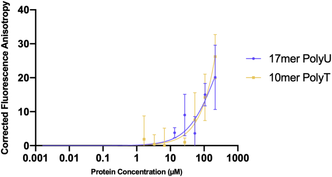
Many of the SARS-CoV-2 proteins have related counterparts across the Severe Acute Respiratory Syndrome (SARS-CoV) family. One such protein is non-structural protein 9 (Nsp9), which is thought to mediate viral replication, overall virulence, and viral genomic RNA reproduction. We sought to better characterize the SARS-CoV-2 Nsp9 and subsequently solved its X-ray crystal structure, in an apo form and, unexpectedly, in a peptide-bound form with a sequence originating from a rhinoviral 3C protease sequence (LEVL). The SARS-CoV-2 Nsp9 structure revealed the high level of structural conservation within the Nsp9 family. The exogenous peptide binding site is close to the dimer interface and impacted the relative juxtapositioning of the monomers within the homodimer. We have established a protocol for the production of SARS-CoV-2 Nsp9, determined its structure, and identified a peptide-binding site that warrants further study to understanding Nsp9 function.

Keywords: 3D Reconstruction of Protein; Protein Structure Aspects; Structural Biology; Virology.


Conflict of interest statement

Declaration of Interests The authors declare no conflict of interest. This article contains Supplemental Information online.

Figures

None
Graphical abstract
Figure 1
Figure 1
Nsp9COV19 Nucleotide-Binding Assay Fluorescence polarization anisotropy assays were used to examine the possibility that Nsp9COV19 could bind to labeled 17-mer and 10-mer single-stranded oligonucleotides. The plot shows corrected anisotropy for each Nsp9COV19 protein concentration; error bars represent the SD from the mean of triplicate measurements after 60 min incubation.
Figure 2
Figure 2
apo-Nsp9COV19 Is Structurally Similar to Nsp9SARS (A–C) Cartoon representation of the monomeric units of (A) apo-Nsp9COV19, (B) apo-Nsp9SARS (Sutton et al., 2004), and (C) a backbone alignment of the two structures. The COV19 structures are colored with β strands in marine and the α helix in wheat; the SARS structures are in teal and orange, respectively. (D) The bound peptide is highlighted in red.
Figure 3
Figure 3
Peptide Binding in Nsp9COV19 Alters the Dimer Interface (A and B) Top-down views of the dimer interface highlighting the interaction helices for (A) unbound Nsp9COV19 in which the surface of the hydrophobic interface cavity is displayed labeled; (B) an equivalent representation of peptide-occupied 3c-Nsp9COV19 dimer. (C) Stick representation of the GxxxG protein-protein interaction helices at the dimer interface for apo-Nsp9COV19. (D) Cα backbone overlay of the Nsp9COV19 interface in the apo and peptide-occupied states. The GxxxG motif residues are colored light purple.
Figure 4
Figure 4
Movements within the GxxxG Motif (A and B) A side view of the N- and C- terminal structural elements at the dimer interface is shown for the (A) apo and (B) peptide-occupied forms. (C) Overlay of the Nsp9COV19 dimer in the apo and peptide-bound forms indicating respective shifts in subunit orientation. The center of mass of the nonaligned subunit is depicted with a light-pink and dark-pink point, respectively. (D) Unbiased omit map contoured at 3.2σ near the hydrophobic cavities into which the exogenous bound peptide was refined.
Figure 5
Figure 5
Sequence Conservation within Nsp9 Homologs (A) Sequence alignment for viral Nsp9 proteins encoded by SARS-CoV-2, SARS-CoV, Human coronavirus 229E, and Porcine epidemic diarrhea virus. The extent of secondary structural elements observed in the 3C-Nsp9COV19 structure is shown and labeled above. The GxxxG motif residues are highlighted with purple and those making up the extraneous peptide binding site in pink. (B) Cartoon-and-stick representation of the peptide-binding site observed in 3C-Nsp9COV19. The side chains of the extraneous 3C residues on one side of the paired helical-interface are displayed with carbon atoms colored pink. Nearby residues that make up the binding site are displayed and labeled and listed in the accompanying contacts table.

References

    1. Chen J., Xu X., Tao H., Li Y., Nan H., Wang Y., Tian M., Chen H. Structural analysis of porcine reproductive and respiratory syndrome virus non-structural protein 7alpha (NSP7alpha) and identification of its interaction with NSP9. Front. Microbiol. 2017;8:853. - PMC - PubMed
    1. Egloff M.P., Ferron F., Campanacci V., Longhi S., Rancurel C., Dutartre H., Snijder E.J., Gorbalenya A.E., Cambillau C., Canard B. The severe acute respiratory syndrome-coronavirus replicative protein nsp9 is a single-stranded RNA-binding subunit unique in the RNA virus world. Proc. Natl. Acad. Sci. U S A. 2004;101:3792–3796. - PMC - PubMed
    1. Frieman M., Yount B., Agnihothram S., Page C., Donaldson E., Roberts A., Vogel L., Woodruff B., Scorpio D., Subbarao K. Molecular determinants of severe acute respiratory syndrome coronavirus pathogenesis and virulence in young and aged mouse models of human disease. J. Virol. 2012;86:884–897. - PMC - PubMed
    1. Hu T., Chen C., Li H., Dou Y., Zhou M., Lu D., Zong Q., Li Y., Yang C., Zhong Z. Structural basis for dimerization and RNA binding of avian infectious bronchitis virus nsp9. Protein Sci. 2017;26:1037–1048. - PMC - PubMed
    1. Miknis Z.J., Donaldson E.F., Umland T.C., Rimmer R.A., Baric R.S., Schultz L.W. Severe acute respiratory syndrome coronavirus nsp9 dimerization is essential for efficient viral growth. J. Virol. 2009;83:3007–3018. - PMC - PubMed

LinkOut - more resources