. 2020 Sep 1;105(9):2983-2995.
doi: 10.1210/clinem/dgaa413.

Human Endometrial Stromal Cell Differentiation is Stimulated by PPARβ/δ Activation: New Targets for Infertility?

Affiliations

Human Endometrial Stromal Cell Differentiation is Stimulated by PPARβ/δ Activation: New Targets for Infertility?

Jie Yu et al. J Clin Endocrinol Metab. .

Abstract

Context: Implantation is a reproductive bottleneck in women, regulated by fluctuations in ovarian steroid hormone concentrations. However, other nuclear receptor ligands are modifiers of endometrial differentiation leading to successful pregnancy. In the present study we analyzed the effects of peroxisome-proliferator-activated receptor β/δ (PPARβ/δ) activation on established cellular biomarkers of human endometrial differentiation (decidualization).

Objective: The objective of this work is to test the effects of PPARβ/δ ligation on human endometrial cell differentiation.

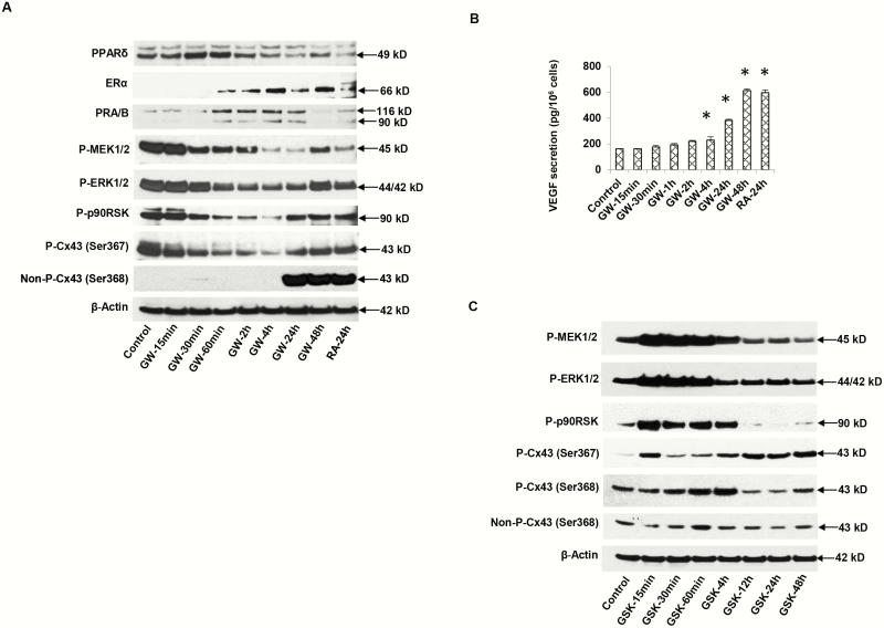
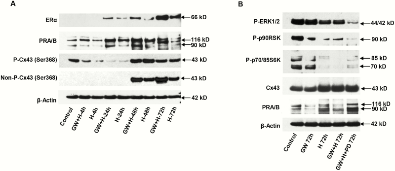
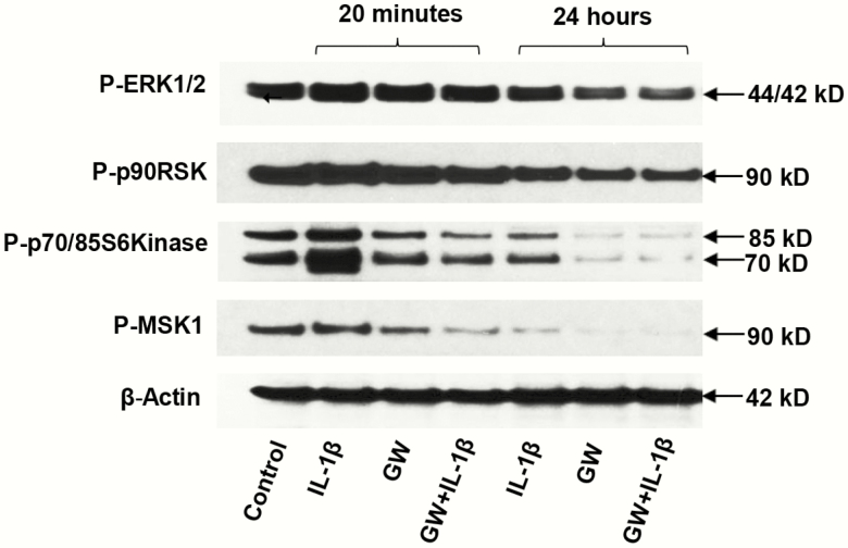
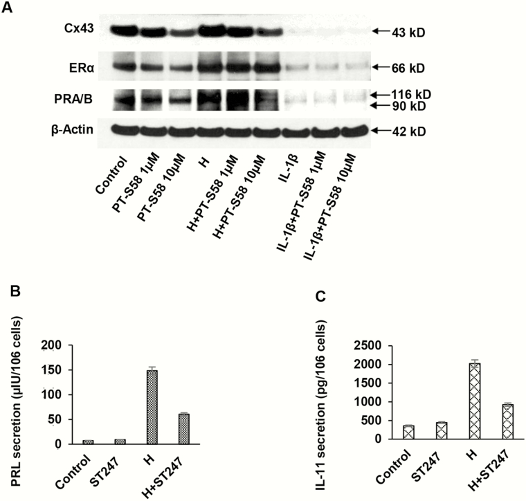
Design: Isolated primary human endometrial stromal cells (ESCs) were treated with synthetic (GW0742) or natural (all trans-retinoic acid, RA) ligands of PPARβ/δ, and also with receptor antagonists (GSK0660, PT-S58, and ST247) in the absence or presence of decidualizing hormones (10 nM estradiol, 100 nM progesterone, and 0.5 mM dibutyryl cAMP [3',5'-cyclic adenosine 5'-monophosphate]). In some cases interleukin (IL)-1β was used as an inflammatory stimulus. Time course and dose-response relationships were evaluated to determine effects on panels of well characterized in vitro biomarkers of decidualization.

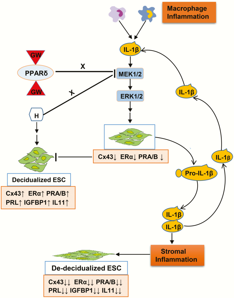
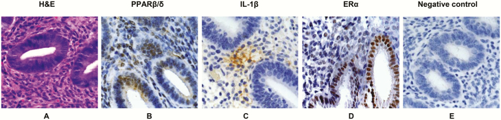
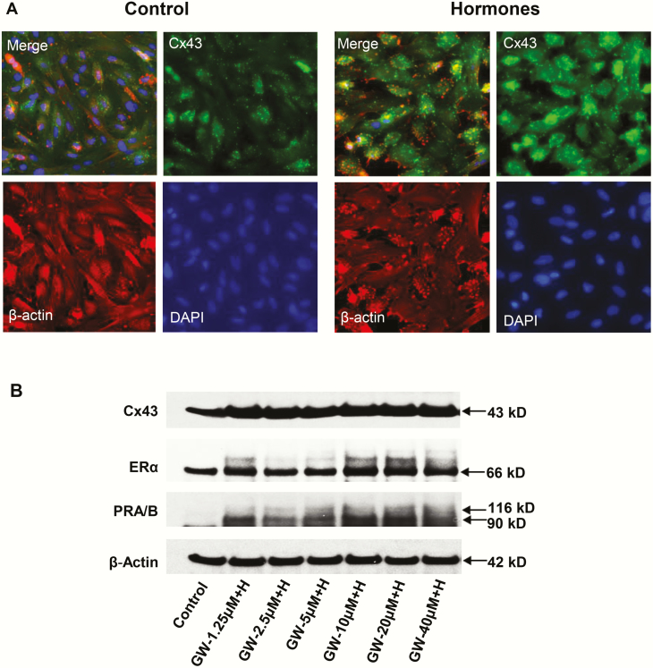
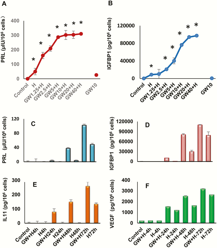
Results: PPARβ/δ, along with estrogen receptor α (ERα) and PR-A and PR-B, were expressed in human endometrial tissue and isolated ESCs. GW0742 treatment enhanced hormone-mediated ESC decidualization in vitro as manifested by upregulation of prolactin, insulin-like growth factor-binding protein 1, IL-11, and vascular endothelial growth factor (VEGF) secretion and also increased expression of ERα, PR-A and PR-B, and connexin 43 (Cx43). RA treatment also increased VEGF, ERα, PR-A, and PR-B and an active, nonphosphorylated isoform of Cx43. IL-1β and PPARβ/δ antagonists inhibited biomarkers of endometrial differentiation.

Conclusion: Ligands that activate PPARβ/δ augment the in vitro expression of biomarkers of ESC decidualization. By contrast, PPARβ/δ antagonists impaired decidualization markers. Drugs activating these receptors may have therapeutic benefits for embryonic implantation.

Keywords: decidualization; fatty acids; nuclear receptors; retinoic acid; uterus.


Figures

Figure 1.
Figure 1.
Graphic model of study hypothesis. Macrophages and other inflammatory cells in the endometrial microenvironment are a primary source of interleukin 1β (IL-1β) and other proinflammatory cytokines, but endometrial stromal cell (ESC) autocrine and paracrine sources also contribute. The major biomarkers evaluated in this report are represented in the pink boxes, with arrows indicating direction and magnitude of production or secretion. Corresponding alterations in ESC shape are represented in the cartoon.
Figure 2.
Figure 2.
Immunohistochemistry of peroxisome-proliferator-activated receptor β/δ (PPARβ/δ), interleukin1β (IL-1β), and estrogen receptor α (ERα) in endometrium. A, A hematoxylin-eosin (H&E)-stained section of proliferative endometrium is provided to illustrate relevant tissue morphology. B, Immunoperoxidase histochemistry (brown precipitate) shows nuclear localization of PPARβ/δ in endometrial glands and stroma. C, Immunoperoxidase histochemistry (brown precipitate) shows focal, lacy stromal cytoplasmic localization of IL-1β in an adjacent section. D, As a positive control, anti-ERα antibodies stained epithelial and stromal nuclei. E, A negative control, in which the primary antibodies were substituted with nonimmune serum. All sections were counterstained with Mayer’s hematoxylin. Similar findings were observed in n = 3 tissue sections. Magnification × 200.
Figure 3.
Figure 3.
A, Time-course of peroxisome-proliferator-activated receptor β/δ (PPARβ/δ) agonist GW0742 (GW) effects on endometrial stromal cell (ESC) protein lysates by Western blotting. GW treatment for 15 minutes to 48 hours showed changes in nuclear receptors: PPARβ/δ (49 kDa, panel 1), estrogen receptor α (ERα) (66 kDa, panel 2), PR-A and -B (90 and 116 kDa, respectively, panel 3). Phosphoproteins of the extracellularly regulated kinase (ERK)1/2 cascade included phospho-MEK1/2 (45 kDa, panel 4), phospho-ERK1/2 (42 kDa, panel 5), and phospho-p90 RSK (90 kDa, panel 6). Connexin 43 (Cx43) isoforms: phospho-Cx43 Ser367 (43 kDa, panel 7), nonphospho-Cx43 Ser368 (43 kDa, panel 8). β-Actin levels (42 kDa, panel 9) are shown. Lane 1 represents untreated control cells and lane 9, ESCs treated for 24 hours with all-trans retinoic acid (RA) as a positive control. B, Under the same conditions, ESC supernatant concentrations of vascular endothelial growth factor (VEGF) were determined by enzyme-linked immunosorbent assay. Means ± SEM of triplicate samples are shown. GW at 24 and 48 hours and RA at 24 hours each significantly stimulated VEGF secretion over control conditions (P < .05, t tests with Bonferroni corrections, n = 3). C, Time-course effects of PPARβ/δ antagonist, GSK0660 (GSK) on ESC protein markers by Western blotting. Phosphoproteins of the extracellularly regulated kinase (ERK)1/2 cascade included phospho-MEK1/2 (45 kDa, panel 1), phospho-ERK1/2 (42 kDa, panel 2), phospho-p90 RSK (90 kDa, panel 3), Cx43 isoforms: Phospho-Cx43 Ser367 (43 kDa, panel 4), phospho-Cx43 Ser368 (43 kDa, panel 5), and nonphospho-Cx43 Ser368 (43 kDa, panel 6). β-Actin levels (42 kDa, panel 7) are shown. Lane 1 represents untreated control cells.
Figure 4.
Figure 4.
Effects of peroxisome-proliferator-activated receptor β/δ (PPARβ/δ) agonist (GW), hormones (H), and MEK inhibitor (PD) were assessed. A, Time-course and additive effects of GW + H for up to 72 hours were observed for endometrial stromal cell (ESC) expression of estrogen receptor α (ERα) (panel 1), PR-A and -B (panel 2), nonphospho-connexin 43 (Cx43) Ser368 (panel 4), and phospho-Cx43 Ser368 (panel 3). β-Actin levels (panel 5) are shown. Lane 1 represents untreated control cells. B, Relative to controls, a combination of GW, H, and PD for 72 hours led to the lowest phospho-extracellularly regulated kinase (ERK)1/2 (panel 1), phospho-p90 RSK (panel 2), phospho-p70/85 S6Kinase (panel 3) and was associated with the highest levels of total Cx43 (panel 4) and PR-A and -B (panel 5) (P < .05, t-test, n = 3). β-Actin levels (panel 6) are shown as loading controls.
Figure 5.
Figure 5.
A, The effects of hormone (“H”) treatment on immunofluorescent connexin 43 (Cx43) (green) staining in endometrial stromal cell (ESC) cultures for 72 hours are shown in the upper right panels. A 2.6 ± 0.5-fold increase in Cx43 pixel count (P < .05, t test, n = 4) was noted. DAPI (4’,6-diamidino-2-phenylindole) nuclear staining (lower right panels), β-actin (lower left panels), and a merged frame (upper left panels) also are presented. B, Western blot results in ESCs treated with different concentrations of peroxisome-proliferator-activated receptor β/δ (PPARβ/δ) agonist (GW) combined with decidualizing hormones (“H”) for Cx43, estrogen receptor α (ERα) and PR-A and PR-B are shown. Standard E2 + P4 + 3′,5′–cyclic adenosine 5′-monophosphate (cAMP) hormone concentrations were kept constant while GW levels were increased. ERα (panel 2) and PR-A and -B (panel 3) both revealed dose-dependent increases by the addition of GW (“GW + H”).
Figure 6.
Figure 6.
Dose-responsive effects of peroxisome-proliferator-activated receptor β/δ (PPARβ/δ) agonist (GW) and hormones (“H”) alone and in combination and time-course effects (GW + H) on A, prolactin (PRL) and B, insulin-like growth factor-binding protein 1 (IGFBP-1) secretion were evaluated by enzyme-linked immunosorbent assay (ELISA). In the presence of H, GW had apparent EC50s ranging from 1 to 5 μM. The incubation time was 72 hours. Asterisks signify dose-response effects different from control conditions (P < .05, n = 3).Time-course effects of GW on secretory behavior of differentiated endometrial stromal cells. ELISAs were used to quantify C, PRL, D, IGFBP-1, E, interleukin 11 (IL-11), and F, vascular endothelial growth factor (VEGF) secretion by these cells. The data reflect the effects of up to 72 hours of H alone, and also the additive responses to a combination of 10 μM GW + H.
Figure 7.
Figure 7.
Effects of peroxisome-proliferator-activated receptor β/δ (PPARβ/δ) agonist (GW) on interleukin 1β (IL-1β)-mediated inhibition of decidualization biomarkers in endometrial stromal cells (ESCs). ESCs were exposed briefly (20 minutes) or chronically (24 hours) to IL-1β, GW, or a combination of the compounds. Phospho (P)-extracellularly regulated kinase (ERK)1/2, P-p90 RSK, P-p70/85 S6K, and P-MSK1 were all significantly reduced by the combination after 24 hours (panels 1-4, respectively) (≤ 0.4 ± 0.1-fold, P < .05, t test, n = 3). β-Actin levels did not change in response to the treatments (panel 5).
Figure 8.
Figure 8.
Alternative peroxisome-proliferator-activated receptor β/δ (PPARβ/δ) inhibitors (PT-S58 and ST247) were used to assess effects on endometrial stromal cell (ESC) biomarkers. A, After a 72-hour incubation period, PT-S58 appeared to dose-dependently suppress basal connexin 43 (Cx43) (panel 1) and basal and hormone-induced estrogen receptor α (ERα) and PR (panels 2 and 3). Interleukin 1β (IL-1β) strongly inhibited Cx43, ERα, and PR to such an extent that it is difficult to ascertain whether the PPARβ/δ antagonist caused further inhibition. At the 10-μM concentration, PT-S58 suppressed basal Cx43 to 0.5 ± 0.1-fold and hormone-stimulated Cx43 to 0.3 ± 0.1-fold control levels (P < .05, t test, n = 3). β-Actin levels were not affected (panel 4). The inverse PPARβ/δ agonist, ST247 (10 μM), was incubated with ESCs for 72 hours in the absence or presence of H. Hormone-induced B, prolactin and C, IL-11 secretion were both inhibited more than 50% by ST247, although the inverse agonist had no effect under control conditions (P < .05, t test, n = 3).

References

    1. Bellofiore N, Ellery SJ, Mamrot J, Walker DW, Temple-Smith P, Dickinson H. First evidence of a menstruating rodent: the spiny mouse (Acomys cahirinus). Am J Obstet Gynecol. 2017;216(1):40.e1-40.e11. - PubMed
    1. Gellersen B, Brosens JJ. Cyclic decidualization of the human endometrium in reproductive health and failure. Endocr Rev. 2014;35(6):851-905. - PubMed
    1. Lessey BA, Young SL. What exactly is endometrial receptivity? Fertil Steril. 2019;111(4):611-617. - PubMed
    1. Devroey P, Pados G. Preparation of endometrium for egg donation. Hum Reprod Update. 1998;4(6):856-861. - PubMed
    1. Shifren JL, Tseng JF, Zaloudek CJ, et al. . Ovarian steroid regulation of vascular endothelial growth factor in the human endometrium: implications for angiogenesis during the menstrual cycle and in the pathogenesis of endometriosis. J Clin Endocrinol Metab. 1996;81(8):3112-3118. - PubMed

Publication types

MeSH terms