. 2020 Jun 29;13(1):99.
doi: 10.1186/s13041-020-00605-5.

Expression of serotonin 1A and 2A receptors in molecular- and projection-defined neurons of the mouse insular cortex

Affiliations

Expression of serotonin 1A and 2A receptors in molecular- and projection-defined neurons of the mouse insular cortex

Anes Ju et al. Mol Brain. .

Abstract

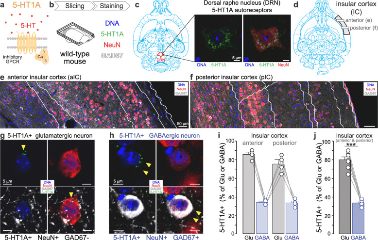
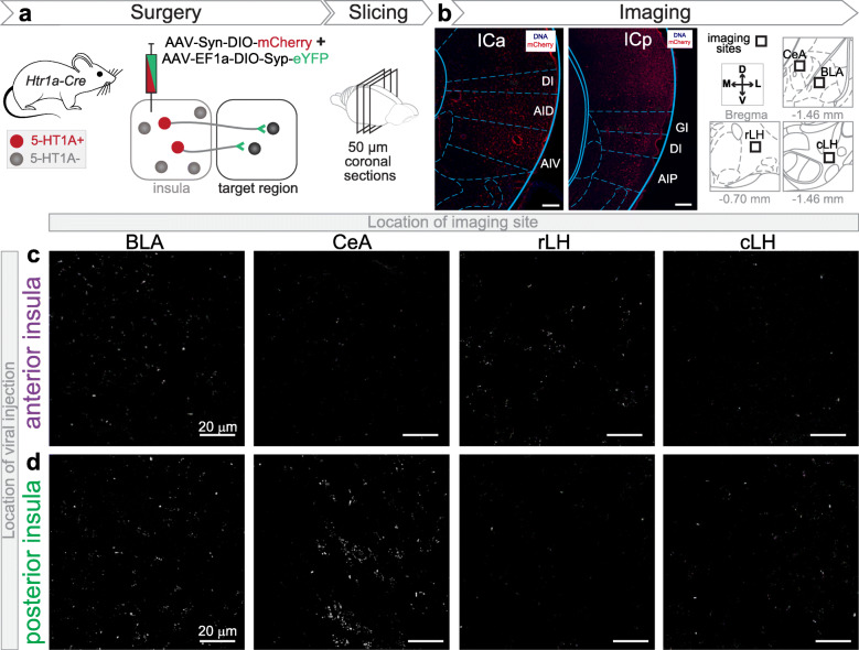
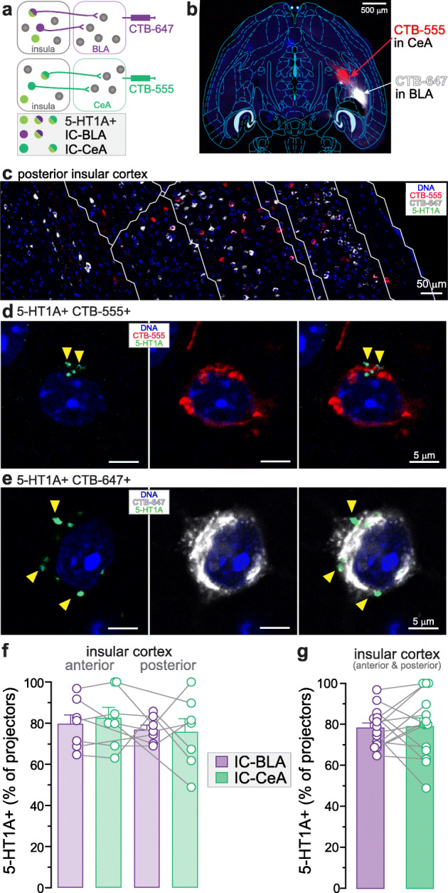
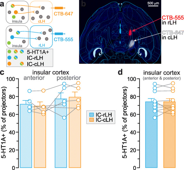
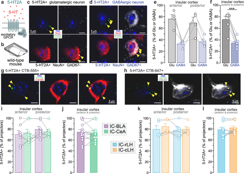
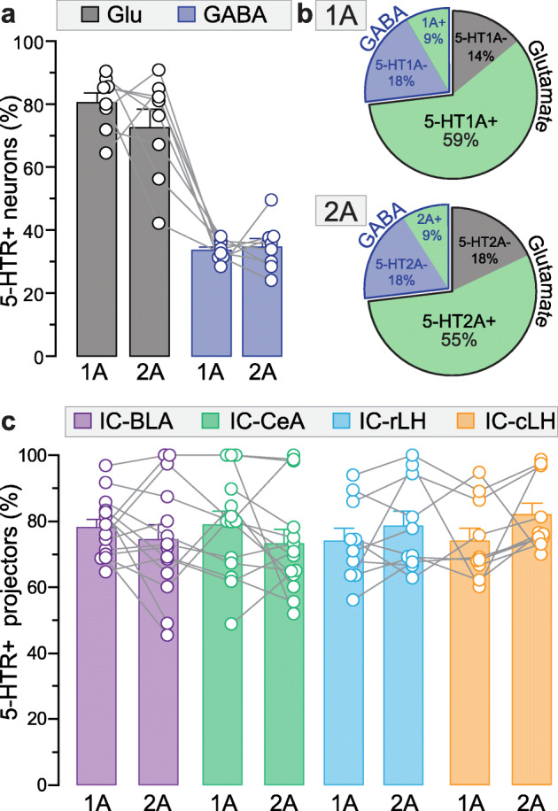
The serotonin (5-HT) system is the target of multiple anxiolytics, including Buspirone, which is a partial agonist of the serotonin 1A receptor (5-HT1A). Similarly, ligands of the serotonin 2A receptor (5-HT2A) were shown to alter anxiety level. The 5-HT1A and 2A receptors are widely expressed across the brain, but the target region(s) underlying the influence of those receptors on anxiety remain unknown. Interestingly, recent studies in human and non-human primates have shown that the 5-HT1A and 5-HT2A binding potentials within the insular cortex (insula) are correlated to anxiety. As an initial step to define the function of 5-HT transmission in the insula, we quantified the proportion of specific neuronal populations of the insula expressing 5-HT1A or 5-HT2A. We analyzed seven neural populations, including three defined by a molecular marker (putative glutamate, GABA or parvalbumin), and four defined by their projections to different downstream targets. First, we found that more than 70% of putative glutamatergic neurons, and only 30% of GABAergic neurons express the 5-HT1A. Second, within insular projection neurons, 5-HT1A is highly expressed (75-80%) in the populations targeting one sub-nuclei of the amygdala (central or basolateral), or targeting the rostral or caudal sections of the lateral hypothalamus (LH). Similarly, 70% of putative glutamatergic neurons and only 30% of insular GABAergic neurons contain 5-HT2A. Finally, the 5-HT2A is present in a majority of insula-amygdala and insula-LH projection neurons (73-82%). These observations suggest that most glutamatergic neurons can respond to 5-HT through 5-HT1A or 5-HT2A in the insula, and that 5-HT directly affects a limited number of GABAergic neurons. This study defines a molecular and neuroanatomical map of the 5-HT system within the insular cortex, providing ground knowledge to identify the potential role of serotonergic modulation of selective insular populations in anxiety.

Keywords: Anxiety; Excitatory neurons; Immunohistochemistry; Inhibitory neurons; Neural circuit; Neuroanatomy; Projection neurons; Retrograde tracing; Synaptophysin.


Conflict of interest statement

The authors declare no competing interests.

Figures

Fig. 1
Fig. 1
Expression of 5-HT1A in the glutamatergic and GABAergic neurons of the insular cortex. a. Molecular diagram of 5-HT1A coupled to an inhibitory G protein. b. Experimental design to identify 5-HT1A-expressing (5-HT1A+) glutamatergic and GABAergic neurons in the insula using immunofluorescent staining in horizontal brain slice of wild-type mouse. c. 5-HT1A antibody staining of soma 5-HT1A-autoreceptors in neurons of the dorsal raphe nucleus (DRN). d. Definition of the analyzed area in the anterior and posterior insula in horizontal brain slices (3.60 mm ventral to Bregma). e-f. Image of DNA staining and immunofluorescent signal of NeuN and GAD67 in the anterior (e) and posterior (f) insula. White lines indicate the division of cortical layers in the insula. g-h. Representative images of 5-HT1A+ glutamatergic (g) and GABAergic neurons (h) in the insula. Yellow arrows indicate 5-HT1A expression. i. Percentage of 5-HT1A expression in glutamatergic (Glu) and GABAergic (GABA) neurons in the anterior (ICa) and posterior insula (ICp). Total glutamatergic neurons: ICa + ICp = 1966 + 2359 = 4325; Total GABAergic neurons: ICa + ICp = 513 + 373 = 886; n = 4 mice j. The proportion of 5-HT1A+ neurons is significantly higher in the glutamatergic than in the GABAergic population (paired t-test, ***p < 0.001), n = 4 mice, 2 locations per mouse
Fig. 2
Fig. 2
Evidence of synaptic contacts of 5-HT1A+ insular neurons in the amygdala and lateral hypothalamus. a. Experimental design to detect synaptic terminals of 5-HT1A-expressing (5-HT1A+) insula neurons through cre-dependent expression of mCherry and synaptophysin-eYFP in the anterior or posterior insula of Htr1a-Cre mice. b. (Left) Confocal image of the cre-dependent viral vector injection site in a coronal section of the anterior (DI: dysgranular insula, AID: agranular insula dorsal part, AIV: agranular insula ventral part) and the posterior insula (GI: granular insula, AIP: agranular insula posterior part). Note mCherry expression in the soma of 5-HT1A+ neurons. (Right) Imaging locations of synaptophysin-eYFP in the basolateral and central amygdala (BLA and CeA) as well as the rostral and caudal part of the lateral hypothalamus (rLH and cLH). Distances are in the anteroposterior axis from Bregma. c-d. Confocal images of the expression of synaptophysin-eYFP in BLA, CeA, rLH and cLH, originating from 5-HT1A+ neurons of the anterior (c) and posterior (d) insula
Fig. 3
Fig. 3
Expression of 5-HT1A in insula neurons projecting to subnuclei of the amygdala. a. Experimental timeline combining retrograde fluorescent tracers (CTB) injections and 5-HT1A immunostaining. b. Injection site of retrograde fluorescent tracers (CTB-555 and CTB-647) in the central and basolateral amygdala (CeA and BLA) within a horizontal brain slice (4.56 mm ventral to Bregma). c. Immunofluorescent image showing CTB-555 and CTB-647 positive neurons in the posterior insula. d-e. Representative fluorescent images of CTB-labelled neurons expressing 5-HT1A in the insula. Yellow arrows indicate 5-HT1A expression. f. Percentage of 5-HT1A expression in neurons projecting to the BLA and CeA in the anterior (ICa) and posterior insula (ICp). g. The proportion of 5-HT1A+ neurons is not different between IC-BLA and IC-CeA populations (unpaired t-test, p > 0.05). Total IC-BLA neurons: ICa + ICp = 417 + 620 = 1037; Total IC-CeA neurons: ICa + ICp = 455 + 647 = 1102; n = 7 mice, 2 locations per mouse
Fig. 4
Fig. 4
Expression of 5-HT1A in insular neurons projecting to the lateral hypothalamus. a. Experimental design combining retrograde fluorescent tracers (CTB) and serotonin 1A receptor (5-HT1A) antibody staining. b. Injection site of retrograde fluorescent tracers (CTB-555 and CTB-647) in the rostral and caudal parts of lateral hypothalamus (rLH and cLH, respectively) within a horizontal brain slice (4.56 mm ventral to Bregma). c. Percentage of 5-HT1A expression in neurons projecting to cLH and rLH from the anterior (ICa) and posterior (ICp) insula. d. The proportion of 5-HT1A+ neurons are not different between IC-rLH and IC-cLH populations (unpaired t-test, p > 0.05). Total IC-rLH neurons: ICa + ICp = 298 + 313 = 611; Total IC-CeA neurons: ICa + ICp = 422 + 244 = 666; n = 5 mice, 2 locations per mouse
Fig. 5
Fig. 5
Expression of 5-HT2A in glutamatergic, GABAergic and projection-specific populations of the insular cortex. a. Molecular diagram of the serotonin 2A receptor (5-HT2A) coupled to an excitatory G-protein. b. Horizontal brain slice of wild-type mouse used in immunofluorescent staining. c-d. Representative images of 5-HT2A-expressing (5-HT2A+) glutamatergic (c) and GABAergic neurons (d) in the insula. Yellow arrows indicate 5-HT2A expression. e. Percentage of 5-HT2A expression in the glutamatergic (Glu) and GABAergic (GABA) neurons in the anterior (ICa) and posterior (ICp) insula. f. The proportion of 5-HT2A+ neurons is significantly higher in glutamatergic than GABAergic populations (paired t-test, ***p < 0.001). Total glutamatergic neurons: ICa + ICp = 2444 + 2783 = 5227; Total GABAergic neurons: ICa + ICp = 542 + 577 = 1119; n = 4 mice. g-h. Representative fluorescent images of CTB-labelled neurons (IC-BLA and IC-CeA projection neurons) expressing 5-HT2A in the insula. Yellow arrows indicate 5-HT2A expression. i. Percentage of 5-HT2A expression in neurons projecting to the central and basolateral amygdala (CeA and BLA) in the anterior and posterior insula. j. The proportions of 5-HT2A+ neurons are not different between IC-BLA and IC-CeA populations (unpaired t-test, p > 0.05). Total IC-BLA neurons: ICa + ICp = 424 + 324 = 748; Total IC-CeA neurons: ICa + ICp = 526 + 523 = 1049; n = 7 mice, 2 locations per mouse. k. The percentage of 5-HT2A expression in neurons projecting to rostral and caudal parts of the lateral hypothalamus (rLH and cLH) in the anterior and posterior insula. l. The proportions of 5-HT2A+ neurons are not different between IC-rLH and IC-cLH populations (unpaired t-test, p > 0.05). Total IC-rLH neurons: ICa + ICp = 530 + 330 = 860; Total IC-CeA neurons: ICa + ICp = 563 + 259 = 822; n = 5 mice, 2 locations per mouse
Fig. 6
Fig. 6
Summary of the proportions of 5-HT1A and 5-HT2A positive neurons in six populations of the insular cortex. a. Percentage of glutamatergic (Glu) and GABAergic (GABA) neurons expressing 5-HT1A or 5-HT2A in the insula (data of Fig. 1j and 5f; 1AR: Glu: 4325 cells, GABA: 886 cells; 2AR: Glu: 5227 cells, GABA: 1119 cells, n = 4 mice; 2 locations per mouse). b. Percentage of glutamatergic and GABAergic neurons containing, or not containing, 5-HT1A (upper) or 5-HT2A (lower) among total insula neurons. c. Percentage of 5-HT1A or 5-HT2A-expressing insular neurons projecting to the basolateral amygdala (IC-BLA), central amygdala (IC-CeA), and rostral or caudal parts of lateral hypothalamus (IC-rLH and IC-cLH, data of Fig. 3g, 4d, 5j and l; 1AR: IC-BLA: 1037 cells, IC-CeA: 1102 cells, n = 7 mice, IC-rLH: 611 cells, IC-cLH: 666 cells, n = 5 mice; 2AR: IC-BLA: 748 cells, IC-CeA: 1049 cells, n = 7 mice, IC-rLH: 860 cells, IC-cLH: 822 cells, n = 5 mice; 2 locations per mouse)

Similar articles

Cited by

References

    1. Baldwin D, Woods R, Lawson R, Taylor D. Efficacy of drug treatments for generalised anxiety disorder: systematic review and meta-analysis. BMJ. 2011;342:d1199. - PubMed
    1. Craske MG, Stein MB. Anxiety. Lancet. 2016;388:3048–3059. - PubMed
    1. Craske MG, Stein MB, Eley TC, Milad MR, Holmes A, Rapee RM, et al. Anxiety disorders. Nature Reviews Disease Primers. 2017;3:17024. - PMC - PubMed
    1. Griebel G, Holmes A. 50 years of hurdles and hope in anxiolytic drug discovery. Nat Rev Drug Discov. 2013;12:667–687. - PMC - PubMed
    1. Tyrer P, Baldwin D. Generalised anxiety disorder. Lancet. 2006;368:2156–2166. - PubMed

Publication types