

Hemoglobin S/OArab: Retinal Manifestations of a Rare Hemoglobinopathy
- PMID: 32595482
- PMCID: PMC7315179
- DOI: 10.1159/000507879
Hemoglobin S/OArab: Retinal Manifestations of a Rare Hemoglobinopathy
Abstract
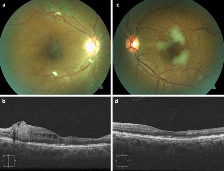
Hemoglobin S/OArab (Hgb S/OArab) disease is a rare hemoglobinopathy which presents similarly to sickle cell retinopathy, with only three prior reports that describe associated retinal findings. In this report, we present ophthalmic examination findings in 2 patients with Hgb S/OArab. One patient exhibited peripheral ischemia and sunburst lesions without neovascular disease, and the other patient developed proliferative retinopathy of both eyes and multiple posterior-pole branch retinal artery occlusions in one eye. To our knowledge, this is the first case of retinal arterial occlusive disease in Hgb S/OArab, and the first report of fundus autofluorescence and OCT angiography in Hgb/OArab retinopathy.
Keywords: Hemoglobin S/OArab; Hemoglobinopathy; Retinal ischemia; Sickle cell retinopathy.
Copyright © 2020 by S. Karger AG, Basel.
Conflict of interest statement
The authors have no conflicts of interest to declare.
Figures


References
-
- Zimmerman SA, O'Branski EE, Rosse WF, Ware RE. Hemoglobin S/O(Arab): thirteen new cases and review of the literature. Am J Hematol. 1999 Apr;60((4)):279–84. - PubMed
-
- Daniel YA, Turner C, Haynes RM, Hunt BJ, Dalton RN. Rapid and specific detection of clinically significant haemoglobinopathies using electrospray mass spectrometry-mass spectrometry. Br J Haematol. 2005 Aug;130((4)):635–43. - PubMed
-
- Zacharia G, Maronge GF, Brazda FW, Boulmay BC. Hemoglobin SO-Arab and α-thalassemia diagnosed in an adult: A case-based review of the hemoglobinopathies. Am J Med Sci. 2013 Oct;346((4)):325–7. - PubMed
-
- Milner PF, Miller C, Grey R, Seakins M, DeJong WW, Went LN. Hemoglobin O arab in four negro families and its interaction with hemoglobin S and hemoglobin C. N Engl J Med. 1970 Dec;283((26)):1417–25. - PubMed
Publication types
LinkOut - more resources
Full Text Sources
