. 2020 Jun 12:11:493.
doi: 10.3389/fphys.2020.00493. eCollection 2020.

Inhibition of Adenosine Pathway Alters Atrial Electrophysiology and Prevents Atrial Fibrillation

Affiliations

Inhibition of Adenosine Pathway Alters Atrial Electrophysiology and Prevents Atrial Fibrillation

Luca Soattin et al. Front Physiol. .

Abstract

Background: Adenosine leads to atrial action potential (AP) shortening through activation of adenosine 1 receptors (A1-R) and subsequent opening of G-protein-coupled inwardly rectifying K+ channels. Extracellular production of adenosine is drastically increased during stress and ischemia.

Objective: The aim of this study was to address whether the pharmacological blockade of endogenous production of adenosine and of its signaling prevents atrial fibrillation (AF).

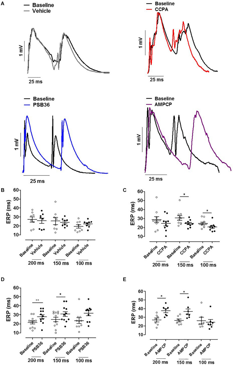
Methods: The role of A1-R activation on atrial action potential duration, refractoriness, and AF vulnerability was investigated in rat isolated beating heart preparations (Langendorff) with an A1-R agonist [2-chloro-N 6-cyclopentyladenosine (CCPA), 50 nM] and antagonist [1-butyl-3-(3-hydroxypropyl)-8-(3-noradamantyl)xanthine (PSB36), 40 nM]. Furthermore, to interfere with the endogenous adenosine release, the ecto-5'-nucleotidase (CD73) inhibitor was applied [5'-(α,β-methylene) diphosphate sodium salt (AMPCP), 500 μM]. Isolated trabeculae from human right atrial appendages (hRAAs) were used for comparison.

Results: As expected, CCPA shortened AP duration at 90% of repolarization (APD90) and effective refractory period (ERP) in rat atria. PSB36 prolonged APD90 and ERP in rat atria, and CD73 inhibition with AMPCP prolonged ERP in rats, confirming that endogenously produced amount of adenosine is sufficiently high to alter atrial electrophysiology. In human atrial appendages, CCPA shortened APD90, while PSB36 prolonged it. Rat hearts treated with CCPA are prone to AF. In contrast, PSB36 and AMPCP prevented AF events and reduced AF duration (vehicle, 11.5 ± 2.6 s; CCPA, 40.6 ± 16.1 s; PSB36, 6.5 ± 3.7 s; AMPCP, 3.0 ± 1.4 s; P < 0.0001).

Conclusion: A1-R activation by intrinsic adenosine release alters atrial electrophysiology and promotes AF. Inhibition of adenosine pathway protects atria from arrhythmic events.

Keywords: A1-R; CD73; adenosine; arrhythmias; hypoxia; translational models.


Figures

FIGURE 1
FIGURE 1
(A) Adenosine (Ado) pathway in cardiomyocytes. Adenosine release is regulated by the ectonucleotidases CD39 and CD73. CD39 converts ATP and ADP into AMP, while CD73 converts AMP into adenosine. Extracellular adenosine half-life is determined by adenosine deaminase (ADA) conversion to inosine (Ino) or internalization by specific equilibrative nucleoside transporters (ENT). A1-receptor activation increases K+ permeability for the G-protein-coupled inwardly rectifying K+ (GIRK) channel through the Gβγ and inhibits the adenylyl cyclase (AC) through Giα subunit. 2-Chloro-N6-cyclopentyladenosine (CCPA) is a potent A1-R agonist; 1-butyl-3-(3-hydroxypropyl)-8-(3-noradamantyl)xanthine (PSB36) specifically antagonizes A1-R signaling. 5′-(α,β-methylene) diphosphate sodium salt (AMPCP) inhibits CD73. (B) Experimental protocol on isolated perfused male Wistar rat hearts. Hearts were removed and placed on the Langendorff configuration. After stabilization phase and baseline, hearts were perfused with vehicle, CCPA, PSB36, or AMPCP, respectively. Each group was paced at CL of 200, 150, and 100 ms. At the end of each experiment, the hearts were exposed to high-frequency pacing events in order to induce AF. (C) Experimental protocol on intact contracting muscles from human right atrial appendages (hRAAs). Human atrial tissues were paced at CL of 1.0 s. After stabilization phase, baseline was recorded for 5 min. After that, hRRAs were exposed to CCPA or PSB36, respectively, for 10 min.
FIGURE 2
FIGURE 2
Chronotropic and dromotropic effects in rat hearts. (A) ECG lead II recordings under intrinsic rhythm. The R–R interval has been used to calculate heart rate. Paired t-tests between the baseline and the drugs were performed to evaluate significant differences. (B) Chronotropic effect of vehicle, 2-chloro-N6-cyclopentyladenosine (CCPA), 1-butyl-3-(3-hydroxypropyl)-8-(3-noradamantyl)xanthine (PSB36), and 5′-(α,β-methylene) diphosphate sodium salt (AMPCP) (gray, red, blue, and purple dot plots, respectively). White dot plots represent the baseline. Values are presented as mean ± SEM. (C) Effect of vehicle, CCPA, PSB36, and AMPCP on the diastolic threshold (DT), respectively. (D) Effect of vehicle, CCPA, PSB36, and AMPCP on Wenckebach’s point, respectively. Vehicle did not affect atrio-ventricular node conduction. CCPA significantly increased Wenckebach point. PSB36 slightly reduced Wenckebach’s point. AMPCP had no effect on AV node conduction. *p < 0.05, **p < 0.01, ***p < 0.001.
FIGURE 3
FIGURE 3
(A) Atrial monophasic action potentials (MAPs) paced at cycle length (CL) of 150 ms before and after the administration of vehicle, 2-chloro-N6-cyclopentyladenosine (CCPA), 1-butyl-3-(3-hydroxypropyl)-8-(3-noradamantyl)xanthine (PSB36), or 5′-(α,β-methylene) diphosphate sodium salt (AMPCP); black MAPs refer to the baseline, while gray, red, blue, and purple MAPs represent hearts perfused with vehicle, CCPA, PSB36, and AMPCP, respectively. (B) Effect of vehicle on APD90 at CL of 200, 150, and 100 ms. (C) CCPA shortened APD90 at CL of 200, 150, and 100 ms, respectively. (D) PSB36 showed a prolongation of APD90 at CL of 200, 150, and 100 ms. (E) Effect of AMPCP on APD90 at CL of 200, 150, and 100 ms. *p ¡ 0.05, **p < 0.01, ***p < 0.001.
FIGURE 4
FIGURE 4
(A) Effective refractory period (ERP) monophasic action potentials (MAPs) of baseline and the relative treated hearts are shown superimposed. (B) Effect of vehicle on ERP at cycle length (CL) 0.20, 0.15, and 0.10 s. (C) 2-Chloro-N6-cyclopentyladenosine (CCPA) shortened refractoriness at CL of 200 ms, significantly at CL of 150 and 100 ms. (D) 1-Butyl-3-(3-hydroxypropyl)-8-(3-noradamantyl)xanthine (PSB36) prolonged ERP at CL of 200 and 150 ms. (E) 5′-(α,β-Methylene) diphosphate sodium salt (AMPCP) prolonged ERP at CL of 200 and 150 ms, but not at 100 ms. *p < 0.05, **p < 0.01.
FIGURE 5
FIGURE 5
Induction of atrial fibrillation (AF) through high-frequency electrical pacing. (A) Atrial monophasic action potentials (MAPs) and pacing traces are shown. Vehicle produced short AF events; by contrasts, 2-chloro-N6-cyclopentyladenosine (CCPA) made the heart prone to sustained AF events. 1-Butyl-3-(3-hydroxypropyl)-8-(3-noradamantyl)xanthine (PSB36) and 5′-(α,β-methylene) diphosphate sodium salt (AMPCP) reduced atrial sensitivity to high frequency electrical pacing (Supplementary Material 6; p < 0.0001). (B) Relative cumulative distribution (in%) of AF duration of each group and dot plot of AF duration events on a logarithmic scale, which shows the range of AF duration among groups when events are different from 0 s. Vehicle, CCPA, PSB36, and AMPCP are depicted in black, red, blue, and purple, respectively.
FIGURE 6
FIGURE 6
(A) A1-R activation in human right atrial appendages (hRAAs). APD90 measurements are shown over time when an isolated contracting atrial muscle was superfused with 1 μM 2-chloro-N6-cyclopentyladenosine (CCPA). (B) Baseline and CCPA-treated hRAA action potentials (APs) were superimposed to show the APD shortening due to A1-R activation. hRAA was paced at cycle length (CL) of 1.0 s. (C) CCPA shortened APD90 in hRAAs. (D) A1-R blockade in hRAAs. APD90 measurements are shown over time when an isolated contracting atrial muscle was superfused with 1 μM 1-butyl-3-(3-hydroxypropyl)-8-(3-noradamantyl)xanthine (PSB36). hRAA was paced at CL of 1,000 ms. (E) Baseline and PSB36-treated hRAA APs were superimposed to show the APD prolongation due to A1-R blockade. (F) PSB36 prolonged APD90 in hRAAs. Resting membrane potential (RMP) in representative panels (B,E) was normalized to show change in AP duration. **p < 0.01, ***p < 0.001.

References

    1. Antonioli L., Pacher P., Vizi E. S., Hasko G. (2013). CD39 and CD73 in immunity and inflammation. Trends Mol. Med. 19 355–367. 10.1016/j.molmed.2013.03.005 - DOI - PMC - PubMed
    1. Atienza F., Almendral J., Moreno J., Vaidyanathan R., Talkachou A., Kalifa J., et al. (2006). Activation of inward rectifier potassium channels accelerates atrial fibrillation in humans: evidence for a reentrant mechanism. Circulation 114 2434–2442. 10.1161/circulationaha.106.633735 - DOI - PubMed
    1. Belardinelli L., Isenberg G. (1983). Isolated atrial myocytes: adenosine and acetylcholine increase potassium conductance. Am. J. Physiol. 244 H734–H737. - PubMed
    1. Belardinelli L., Linden J., Berne R. M. (1989). The cardiac effects of adenosine. Prog. Cardiovasc. Dis. 32 73–97. 10.1016/0033-0620(89)90015-7 - DOI - PubMed
    1. Belardinelli L., Shryock J. C., Song Y., Wang D., Srinivas M. (1995). Ionic basis of the electrophysiological actions of adenosine on cardiomyocytes. FASEB J. 9 359–365. 10.1096/fasebj.9.5.7896004 - DOI - PubMed