Review
. 2020 Jun 12:11:1250.
doi: 10.3389/fimmu.2020.01250. eCollection 2020.

To the Skin and Beyond: The Immune Response to African Trypanosomes as They Enter and Exit the Vertebrate Host

Affiliations
Review

To the Skin and Beyond: The Immune Response to African Trypanosomes as They Enter and Exit the Vertebrate Host

Omar A Alfituri et al. Front Immunol. .

Erratum in

Abstract

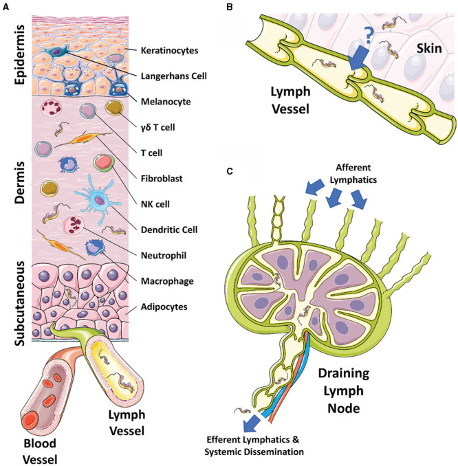
African trypanosomes are single-celled extracellular protozoan parasites transmitted by tsetse fly vectors across sub-Saharan Africa, causing serious disease in both humans and animals. Mammalian infections begin when the tsetse fly penetrates the skin in order to take a blood meal, depositing trypanosomes into the dermal layer. Similarly, onward transmission occurs when differentiated and insect pre-adapted forms are ingested by the fly during a blood meal. Between these transmission steps, trypanosomes access the systemic circulation of the vertebrate host via the skin-draining lymph nodes, disseminating into multiple tissues and organs, and establishing chronic, and long-lasting infections. However, most studies of the immunobiology of African trypanosomes have been conducted under experimental conditions that bypass the skin as a route for systemic dissemination (typically via intraperitoneal or intravenous routes). Therefore, the importance of these initial interactions between trypanosomes and the skin at the site of initial infection, and the implications for these processes in infection establishment, have largely been overlooked. Recent studies have also demonstrated active and complex interactions between the mammalian host and trypanosomes in the skin during initial infection and revealed the skin as an overlooked anatomical reservoir for transmission. This highlights the importance of this organ when investigating the biology of trypanosome infections and the associated immune responses at the initial site of infection. Here, we review the mechanisms involved in establishing African trypanosome infections and potential of the skin as a reservoir, the role of innate immune cells in the skin during initial infection, and the subsequent immune interactions as the parasites migrate from the skin. We suggest that a thorough identification of the mechanisms involved in establishing African trypanosome infections in the skin and their progression through the host is essential for the development of novel approaches to interrupt disease transmission and control these important diseases.

Keywords: African trypanosomiasis; Trypanosoma brucei; innate immunity; neglected tropical disease; skin; transmission.


Figures

Figure 1
Figure 1
The skin, draining lymphatics, and lymph nodes. (A) Diagram of the cellular composition of the epidermal, dermal, and subcutaneous layers of mammalian skin. The outermost epidermal layer consists of a layer of corneocytes above a layer of keratinocytes. These cells manage the tight junctions and the stratum corneum. Langerhans cells and intraepithelial T cells survey the epidermis for antigen to be presented. The central dermal layer contains fibroblasts that produce extracellular matrix proteins to provide structural support and elasticity. Immune responses are initiated by dermal macrophages, dermal dendritic cells, NK cells, and T cells. The inner subcutaneous layer primarily consists of adipocytes. Local lymphatic and blood vessels allow for the trafficking of cells, proteins, and waste. The initial tsetse fly bite injects trypanosomes into the dermis. From the dermis, the parasites exhibit tropism that leads to migration toward the afferent lymph vessels in the skin disseminating to the blood and other regions of the body. (B) The mechanism behind directional migration of trypanosomes from the skin to the lymphatics is unknown. Parasites may be responding to an unreported chemical cue in a chemotactic manner and they may crawl along lymph vessels, access open junctions, or are drawn into the lymphatics through hydrodynamic flow force and pressure. (C) Afferent lymphatic vessels in the skin allow for the drainage of leukocytes and antigen into the draining lymph node. Lymph, containing activated T and B cells, plasma cells, and antibody, passes into the medullary sinus, before exiting via efferent lymphatic vessels. Trypanosomes enter the draining lymph nodes, causing lymphadenopathy, and exit via the efferent lymphatics. Systemic dissemination of the host is reached via the main lymphatic ducts.
Figure 2
Figure 2
The role of innate immune cells during African trypanosomiasis. During early trypanosome infection, a strong Th1 immune response is initiated by the host. In the skin, neutrophils and NK cells are the first to respond to trypanosomal pathogen-associated molecular patterns (PAMPs), such as VSG and CpG DNA. Neutrophils are heavily involved in repairing the initial wound caused by the tsetse fly bite and also produce pro-inflammatory IL-1β and IL-6. NK cells produce pro-inflammatory TNF and IFN-γ that results in the classical activation of pro-inflammatory macrophages (Mθ) via iNOS activation. Macrophages can also be activated through interactions with trypanosomal PAMPs. Classically activated macrophages produce further pro-inflammatory molecules, including TNF, nitric oxide (NO), and reactive nitrogen intermediates (RNIs) and reactive oxygen intermediates (ROIs). These chemicals can directly kill trypanosomes in extravascular spaces and tissues and allows for parasite control. Macrophage secretion of TNF can also recruit and activate T cells which self-renew via autocrine IL-2 secretion. T cells produce IFN-γ to further activate macrophages and IL-4 to activate B cells. Macrophages and dendritic cells (DCs) will further activate B cells during a Th1 response, via IL-6, IL-12, and IFN-γ, to promote the production of antibodies that can target the VSG trypanosomes, inducing waves of parasite clearance in the bloodstream. However, antigenic variation hinders the effective clearance of trypanosome populations. Macrophages can also become alternatively activated, resulting in a Th2 immunosuppressive response. Cytokines such as IL-10, IL-4, and TGF-β initiate this type of response by promoting arginase activation in macrophages. As a result, alternatively activated macrophages produce immunosuppressive IL-10 and suppress production of trypanostatic NO and IFN-γ. This promotes parasite growth and survival, leading to a chronic infection.
Figure 3
Figure 3
The roles of classically and alternatively activated macrophages. (Left) Pro-inflammatory stimuli, such as IFN-γ, TNF-α, and LPS, classically activates macrophages. Classically activated macrophages induce the expression of the inducible nitric oxide synthase (iNOS) enzyme, which catabolizes the substrate L-arginine to produce NO and citrulline. This results in a pro-inflammatory Th1 immune response that can effectively control the initial parasitaemia peak. (Right) Anti-inflammatory stimuli, such as CSF, IL-4, IL-10, IL-13, and TGF-β, alternatively activate macrophages. These induce the expression of the arginase enzyme that catabolizes the substrate L-arginine to produce ornithine. The enzyme ornithine decarboxylase (ODC) catalyzes the breakdown of ornithine to produce polyamines, resulting in an anti-inflammatory Th2 immune response and tissue repair. This type of response typically leads to an immune environment that promotes trypanosome growth and survival.

References

    1. de Raadt P. The history of sleeping sickness. In: Fourth International Course on African Trypanosomiasis. (2005)
    1. Steverding D. The history of African trypanosomiasis. Parasites Vectors. (2008) 1:3. 10.1186/1756-3305-1-3 - DOI - PMC - PubMed
    1. Baral TN. Immunobiology of African trypanosomes: need of alternative interventions. J Biomed Biotechnol. (2010) 2010:389153. 10.1155/2010/389153 - DOI - PMC - PubMed
    1. Morrison LJ, Vezza L, Rowan T, Hope JC. Animal African trypanosomiasis: time to increase focus on clinically relevant parasite and host species. Trends Parasitol. (2016) 32:599–607. 10.1016/j.pt.2016.04.012 - DOI - PubMed
    1. Barrett MP, Burchmore RJ, Stich A, Lazzari JO, Frasch AC, Cazzulo JJ, et al. The trypanosomiases. Lancet. (2003) 362:1469–80. 10.1016/s0140-673614694-6 - DOI - PubMed

Publication types

LinkOut - more resources