. 2020 Jun;15(2):153-162.
doi: 10.1016/j.jds.2020.03.004. Epub 2020 Apr 10.

The advantage of cone-beam computerized tomography over panoramic radiography and temporomandibular joint quadruple radiography in assessing temporomandibular joint osseous degenerative changes

Affiliations

The advantage of cone-beam computerized tomography over panoramic radiography and temporomandibular joint quadruple radiography in assessing temporomandibular joint osseous degenerative changes

Chih-Mong Tsai et al. J Dent Sci. 2020 Jun.

Abstract

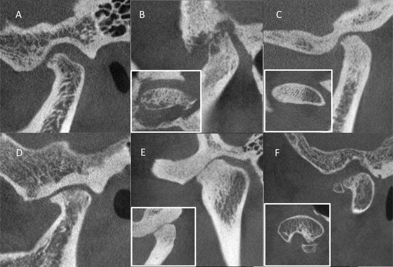
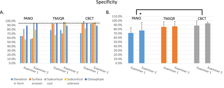
Background/purpose: The clinical diagnosis of temporomandibular joint (TMJ) degenerative joint disease (DJD) is based primarily on radiographic features of the condyle and articular eminence. The purpose of this study was to compare the reliability, sensitivity, and specificity of using plain radiography to that of cone-beam computerized tomography (CBCT) in identifying different types of osseous degenerative features in the TMJ condyle.

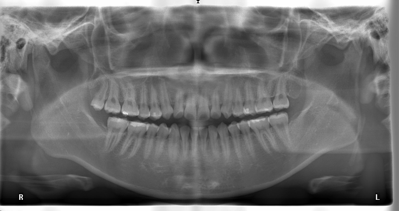
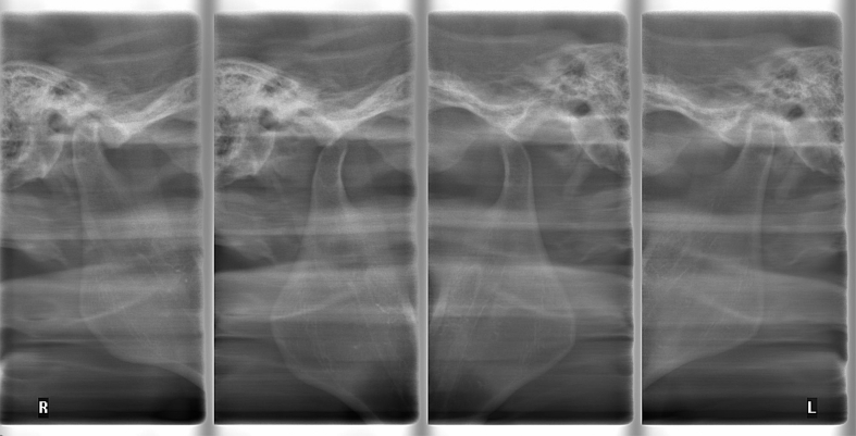
Materials and methods: The panoramic radiography (PANO), TMJ quadruple radiography (TMJQR) and CBCT images of 29 patients' TMJs were retrieved from a computer database and independently evaluated by a young oral surgeon and a senior TMD specialist. The examiners diagnosed osseous degenerative features on the radiographic images. The radiologist-assisted CBCT diagnoses were used as a reference standard and the reliability, sensitivity, and specificity of using the three radiographic modalities were statistically analyzed.

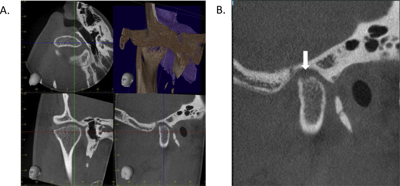
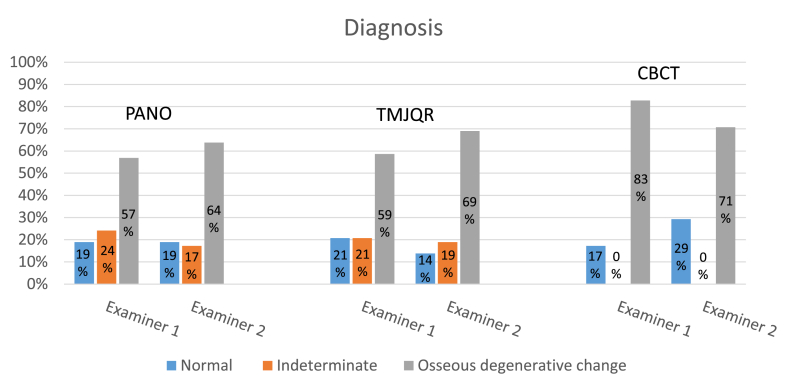
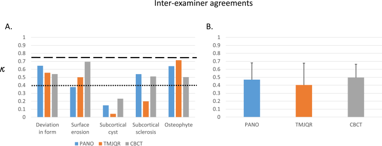
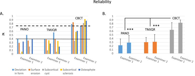
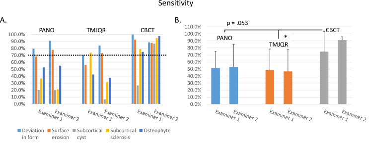
Results: There were cases of indeterminate diagnoses using the PANO and TMJQR due to superimposition from surrounding structures, but none using CBCT. Reliability was generally poor when using PANO and TMJQR for detecting osseous degenerative features of the TMJ condyle but good to excellent when using CBCT. The sensitivity and specificity in the use of PANO and TMJQR were typically below acceptable, but the levels were generally satisfactory when using CBCT.

Conclusion: CBCT is superior to plain radiographic modalities for diagnosing osseous degenerative features of TMJs with regard to indeterminate cases, reliability, sensitivity, and specificity. It is recommended that CBCT can be used as an effective tool in identifying TMJ osteoarthritis.

Keywords: Cone-beam computerized tomography; Osseous degenerative features; Panoramic radiography; TMJ; TMJ quadruple Radiography.


Figures

Figure 1
Figure 1
An example of the panoramic radiograph from one of the patients. It showed the maxilla and mandible, including the dentition and both of the condyles located in the TMJ fossa. In this case, the left and right TMJs were partially superimposed by surrounding bony structures of the cranial base.
Figure 2
Figure 2
The TMJ quadruple radiograph from the same subject presented in Fig. 1. The images at the far left and far right showed the right and left TMJ condyles in the fossa when the mouth was closed; the condyles were superimposed by the surrounding bony structures. The images at the central left and central right showed the condyles out of the fossa when the mouth was widely opened. Even in this case when the TMJs were out of the fossa, it was still not easy for the examiners to judge if there were any osseous degenerative features in the TMJ condyles.
Figure 3
Figure 3
The cone-beam CT images of the right TMJ of the same patient in Figure 1, Figure 2 on the i-Dixel3DXsoftware. A. The volume rendered image (upper-right), the slice images of the axial view (upper-left), the front view (lower-left), and sagittal view (lower-right). The three cursor lines can be moved by use of the computer mouse to implement the slice-by-slice examination of the condyle. B. The magnified sagittal view. The arrow points to a surface erosion that was superposed by surrounding bony structure in the panoramic radiograph image presented in Fig. 1 and could be overlooked by examiners when viewing the TMJ quadruple radiograph in image presented in Fig. 2.
Figure 4
Figure 4
Examples of osseous degenerative features observed in CBCT: A. Deviation in form; B. Subcortical cyst; C. Subcortical sclerosis; D. Osteophyte; E. Bony ankyloses; F. Calcified chondromatosis. Surface erosioncan be seen on the condylar surface in images A, B, C, D, and F.
Figure 5
Figure 5
There are indeterminate cases using the two plain radiographies due to structure superimposition or positioning of the patients; there were no indeterminate cases using CBCT.
Figure 6
Figure 6
A. Inter-examiner agreements (κ) for diagnosis of TMJ degenerative features. The dotted line labels the κ values of0.40 and the dashed line labels the κ values of 0.75. B. Comparison of the inter-examiner agreements among using the three radiographic modalitiesshows no significant difference (p > .05, one-way ANOVA test).
Figure 7
Figure 7
A. Reliability (κ) for diagnosis of TMJ osseous degenerative features using the three modalities by the two examiners. The dotted line labels the κ values of 0.40 and the dashed line labels the κ values of >0.75. B. Comparison of the reliability for identification of TMJ osseous degenerative features using the three modalities by the two examiners. There were significantly higher reliabilities using CBCT than using PANO and TMJQR (∗∗∗p < .001, post-hoc analysis, Bonferronitest).
Figure 8
Figure 8
A. Sensitivity using the three modalities by the two examiners in diagnosing different TMJ osseous degenerative features. The dotted line indicates the acceptable 70% for sensitivity. B. Comparison of the sensitivities for identification of TMJ osseous degenerative features using the three modalities by the two examiners. There was a significantly higher sensitivity using CBCT than using TMJQR (∗p < .05, post-hoc analysis, Bonferronitest) and a nearly significant difference between CBCT and PANO.
Figure 9
Figure 9
A. Specificity using the three modalities by the two examiners in diagnosing different TMJ osseous degenerative features. The dotted line indicates the acceptable 95% for specificity. B. Comparison of the specificities for identification of TMJ osseous degenerative features using the three modalities by the two examiners. Using CBCT had a significantly higher specificity than using PANO (∗p < .05, post-hoc analysis, Bonferronitest).

References

    1. Laskin D., Greenfield W., Gale E. The President's conference on the examination, diagnosis and management of temporomandibular disorders. Chicago: American Dental Association.Kopp S. Topographical distribution of sulfated glycosaminoglycans in the surface layers of the human temporomandibular joint. J Oral Pathol. 1983;7:283–294. - PubMed
    1. Schiffman E., Ohrbach R., Truelove E. Diagnostic criteria for temporomandibular disorders (DC/TMD) for clinical and Research applications: recommendations of the international RDC/TMD consortium network and orofacial pain special interest group. J Oral Facial Pain Headache. 2014;28:6–27. - PMC - PubMed
    1. Embree M., Ono M., Kilts T. Role of subchondral bone during early-stage experimental TMJ osteoarthritis. J Dent Res. 2011;90:1331–1338. - PMC - PubMed
    1. Jiao K., Niu L.N., Wang M.Q. Subchondral bone loss following orthodontically induced cartilage degradation in the mandibular condyles of rats. Bone. 2011;48:362–371. - PubMed
    1. Wang X.D., Kou X.X., He D.Q. Progression of cartilage degradation, bone resorption and pain in rat temporomandibular joint osteoarthritis induced by injection of iodoacetate. PloS One. 2012;7 - PMC - PubMed

LinkOut - more resources