. 2020 Jun 18:8:e9402.
doi: 10.7717/peerj.9402. eCollection 2020.

KIT is involved in melanocyte proliferation, apoptosis and melanogenesis in the Rex Rabbit

Affiliations

KIT is involved in melanocyte proliferation, apoptosis and melanogenesis in the Rex Rabbit

Shuaishuai Hu et al. PeerJ. .

Abstract

Background: Melanocytes play an extremely important role in the process of skin and coat colors in mammals which is regulated by melanin-related genes. Previous studies have demonstrated that KIT is implicated in the process of determining the color of the coat in Rex rabbits. However, the effect of KIT on the proliferation and apoptosis of melanocytes and melanogenesis has not been clarified.

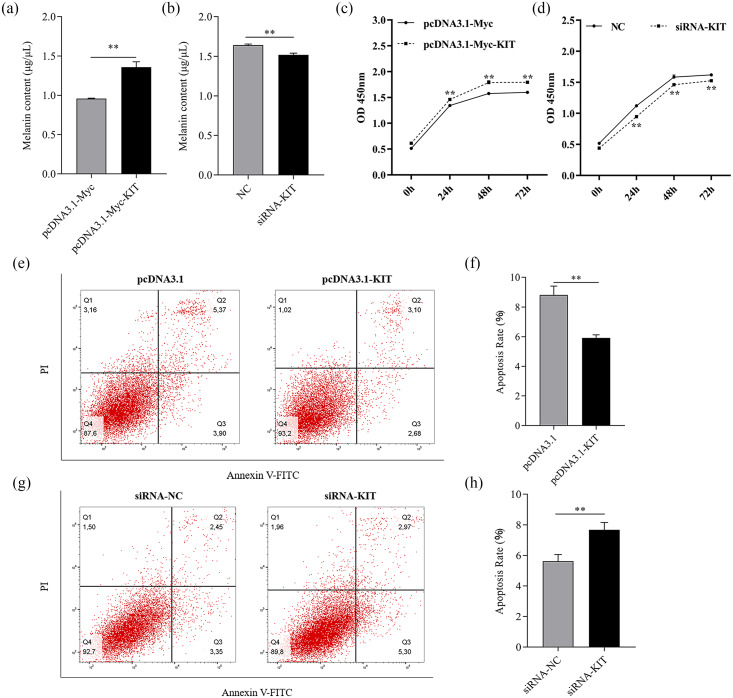
Methods: The mRNA and protein expression levels of KIT were quantified in different coat colored rabbits by qRT-PCR and a Wes assay. To identify whether KIT functions by regulating of melanogenesis, KIT overexpression and knockdown was conducted in melanocytes, and KIT mRNA expression and melanin-related genes TYR, MITF, PMEL and DCT were quantified by qRT-PCR. To further confirm whether KIT influences melanogenesis in melanocytes, melanin content was quantified using NaOH lysis after overexpression and knockdown of KIT. Melanocyte proliferation was estimated using a CCK-8 assay at 0, 24, 48 and 72 h after transfection, and the rate of apoptosis of melanocytes was measured by fluorescence-activated cell sorting.

Results: KITmRNA and protein expression levels were significantly different in the skin of Rex rabbits with different color coats (P < 0.05), the greatest levels observed in those with black skin. The mRNA expression levels of KIT significantly affected the mRNA expression of the pigmentation-related genes TYR, MITF, PMEL and DCT (P < 0.01). Melanin content was evidently regulated by the change in expression patterns of KIT (P < 0.01). In addition, KIT clearly promoted melanocyte proliferation, but inhibited apoptosis.

Conclusions: Our results reveal that KIT is a critical gene in the regulation of melanogenesis, controlling proliferation and apoptosis in melanocytes, providing additional evidence for the mechanism of pigmentation of animal fur.

Keywords: Apoptosis; KIT; Melanocyte; Melanogenesis; Proliferation.


Conflict of interest statement

The authors declare there are no competing interests.

Figures

Figure 1
Figure 1. The Rex rabbits of different coat colors.
(A) White, (B) black, (C) chinchilla, (D) brown, (E) gray, (F) gray-yellow.
Figure 2
Figure 2. Cloning of the KIT gene and mRNA expression in Rex rabbits with different coat colors.
(A–C) 5′ UTR , 3′ UTR and cDNA of KIT was acquired by RACE and cloning techniques. (D) Neighbor-joining phylogenetic tree of Oryctolagus cuniculus and other species based on KIT cDNA. (E) mRNA expression of KIT gene in rabbits of different coat colors by qPCR. (F) KIT protein expression in rabbits of different coat colors was detected by Western blotting. Small letters indicate significant differences among groups (P < 0.05). WH: White, BL: Black, CH: Chinchilla, BR: Brown, GR: Gray, GY: Gray-yellow.
Figure 3
Figure 3. mRNA expression of melanogenesis-related genes was regulated by the change of KIT expression.
(A) The conserved domain of KIT was predicted by NCBI. (B) KIT mRNA expression levels was detected by qPCR after transfection with siRNAs-KIT. (C) mRNA expression of melanogenesis-related genes was analyzed by qPCR after knockdown of KIT. (D) mRNA expression levels of KIT was detected by qPCR when KIT was overexpressed. (E) mRNA expression levels of melanogenesis-related genes as detected by qPCR when KIT was overexpressed. ** P < 0.01, * P < 0.05.
Figure 4
Figure 4. Melanogenesis, melanocyte proliferation and apoptosis were controlled by the change of KIT expression.
(A–B) 48 h after transfection with overexpression or knockdown of KIT, melanin content was analyzed by NaOH lysis. (C–D) Melanocyte proliferation was estimated by CCK-8 assay 24, 48, and 72 h after overexpression or knockdown of KIT. (E–F) Cell apoptosis rate in melanocyte was assessed after overexpression or knockdown of KIT. ** P < 0.01, * P < 0.05.

References

    1. Baxter LL, Watkins-Chow DE, Pavan WJ, Loftus SK. A curated gene list for expanding the horizons of pigmentation biology. Pigment Cell Melanoma Research. 2019;32:348–358. doi: 10.1111/pcmr.12743. - DOI - PMC - PubMed
    1. Bennett DC, Lamoreux ML. The color loci of mice–a genetic century. Pigment Cell Melanoma Research. 2003;16:333–344. doi: 10.1034/j.1600-0749.2003.00067.x. - DOI - PubMed
    1. Brenig B, Beck J, Floren C. Molecular genetics of coat colour variations in White Galloway and White Park cattle. 2013 doi: 10.1111/age.12029. 44:450–453. - DOI - PubMed
    1. Carlie G, Ntusi NBA, Hulley PA, Kidson SH. KUVA (khellin plus ultraviolet A) stimulates proliferation and melanogenesis in normal human melanocytes and melanoma cells in vitro. British Journal of Dermatology. 2015;149:707–717. doi: 10.1046/j.1365-2133.2003.05577.x. - DOI - PubMed
    1. Chen T, Zhao B, Yu L, Wang R, Yang Y, Yang L, Dong CS. MITF-M regulates melanogenesis in mouse melanocytes. Journal of Dermatological Science. 2018;90:253–262. doi: 10.1016/j.jdermsci.2018.02.008. - DOI - PubMed

LinkOut - more resources