

Isolated Primary Rhinosporidiosis of the Parotid Duct: A Rare Presentation
- PMID: 32596180
- PMCID: PMC7302534
- DOI: 10.22038/ijorl.2020.43051.2408
Isolated Primary Rhinosporidiosis of the Parotid Duct: A Rare Presentation
Abstract
Introduction: The primary involvement of the parotid duct in rhinosporidiosis is very rare in clinical practice. Here, we present a case of rhinosporidiosis primarily involving the parotid duct, which was successfully excised through transparotid and transoral approaches.
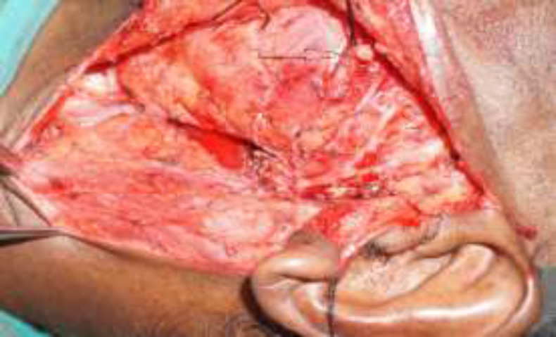
Case report: A 51-year-old male presented with a painless progressive swelling over the left cheek for nine months. It was diagnosed as a parotid cyst or a mucous retention cyst based upon the radiological and cytological features. The cyst was completely excised with transparotid and transoral approaches, and the final diagnosis was confirmed to be rhinosporidiosis.
Conclusion: Although the nose and the paranasal sinus are the common sites to be involved in rhinosporidiosis, the affection of the parotid duct is very unusual in clinical practice.
Keywords: Parotid duct; Primary rhinosporidiosis; Surgical excision.
Figures


References
-
- Arseculeratne SN1. Recent advances in rhinosporidiosis and Rhinosporidium seeberi. Indian J Med Microbiol. 2002;20(3):119–31. - PubMed
-
- Gonazalez G, Viada J, Escalona A, Na'quira N. Nasal rhinosporidiosis - four cases relate literature review. Int Arch Otorhinolaryngol. 2007;11:428–9.
-
- Gonazalez G, Viada J, Escalona A, Na'quira N. Nasal rhinosporidiosis - four cases relate literature review. Int Arch Otorhinolaryngol. 2007;11:428–9.
-
- Babu S, Anuradha A, Chandra S, Kashyap B. Rhinosporidiosis: a case report with review of literature. Ann Trop Med Public Health. 2012;5:127–129.
-
- Sakamoto S, Ogata J, Sakazaki Y, Ikegami K. Fungus ball formation of aspergillus in the bladder An unusual case report. Eur Urol. 1978;4:388–9. - PubMed
Publication types
LinkOut - more resources
Full Text Sources