

Maternal, Fetal, and Placental Selectins in Women With Pre-eclampsia; Association With the Renin-Angiotensin-System
- PMID: 32596247
- PMCID: PMC7304321
- DOI: 10.3389/fmed.2020.00270
Maternal, Fetal, and Placental Selectins in Women With Pre-eclampsia; Association With the Renin-Angiotensin-System
Abstract
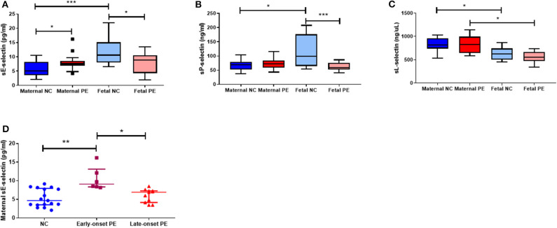
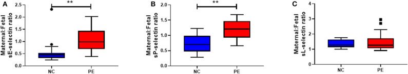
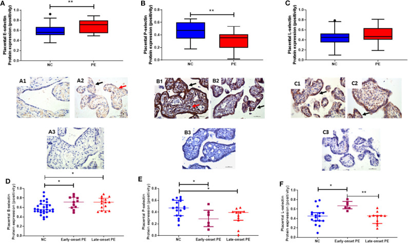
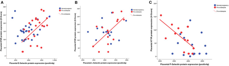
Selectins [endothelial (E), platelet (P), and leucocytes (L)] are a class of cell adhesion molecules, stimulated in response to inflammation. Pre-eclampsia is characterized by inflammation, and angiotensin II is pro-inflammatory. We hypothesized that circulating maternal and fetal concentrations and placental expression of selectins would be increased in women with pre-eclampsia and would be associated with the angiotensin receptors (AT1R and AT2R). Maternal and fetal blood and placental tissue was collected at delivery from White European normotensive controls (n = 17) and women with pre-eclampsia (n = 17). Soluble (s) E-, P- and L-selectin protein concentrations were measured by ELISA and placental protein expression was examined by immunohistochemistry. Maternal sE-selectin concentrations were increased in pre-eclampsia (P < 0.001); conversely fetal sE- and sP-selectin levels were lower in pre-eclampsia (P < 0.05 for both). Staining was mainly localized to the syncytiotrophoblast for all selectins. E-selectin expression was increased, while P-selectin was decreased in placental from pre-eclampsia (P < 0.05 for both); no differences were observed for L-selectin expression. Both E- and L-selectin were positively correlated (P < 0.008; P < 0.02) with AT2R placental expression, whilst P-selectin was negatively associated with AT1R (P < 0.005), all only in the pre-eclampsia group. This novel study reports maternal, fetal and placental expression of selectins in pre-eclampsia. The increased E-selectins reflect the endothelial dysfunction, characteristic of pre-eclampsia. In contrast, the reduced P-selectins and the positive association of placental AT2Rs with both E-and L-selectin in pre-eclampsia could be a protective mechanism to limit the endothelial dysfunction.
Keywords: angiotensin receptors; endothelial dysfunction; inflammation; pre-eclampsia; selectins.
Copyright © 2020 Mistry, Ogalde, Broughton Pipkin, Escher and Kurlak.
Figures
Similar articles
-
The placental renin-angiotensin system and oxidative stress in pre-eclampsia.Placenta. 2013 Feb;34(2):182-6. doi: 10.1016/j.placenta.2012.11.027. Epub 2012 Dec 14. Placenta. 2013. PMID: 23246097
-
Soluble adhesion molecule profile in normal pregnancy and pre-eclampsia.J Matern Fetal Neonatal Med. 2002 Jul;12(1):19-27. doi: 10.1080/jmf.12.1.19.27. J Matern Fetal Neonatal Med. 2002. PMID: 12422905
-
Plasma soluble endothelial selectin is elevated in women with pre-eclampsia.Hum Reprod. 1998 Dec;13(12):3537-41. doi: 10.1093/humrep/13.12.3537. Hum Reprod. 1998. PMID: 9886546
-
Importance of the (Pro)renin Receptor in Activating the Renin-Angiotensin System During Normotensive and Preeclamptic Pregnancies.Curr Hypertens Rep. 2024 Dec;26(12):483-495. doi: 10.1007/s11906-024-01316-1. Epub 2024 Aug 2. Curr Hypertens Rep. 2024. PMID: 39093387 Free PMC article. Review.
-
Potential cell signalling mechanisms involved in differential placental angiogenesis in mild and severe pre-eclampsia.Curr Vasc Pharmacol. 2009 Oct;7(4):475-85. doi: 10.2174/157016109789043865. Curr Vasc Pharmacol. 2009. PMID: 19485894 Review.
Cited by
-
Diagnostic biomolecules and combination therapy for pre-eclampsia.Reprod Biol Endocrinol. 2022 Sep 6;20(1):136. doi: 10.1186/s12958-022-01003-3. Reprod Biol Endocrinol. 2022. PMID: 36068569 Free PMC article. Review.
-
Endothelial Dysfunction in Pregnancy Complications.Biomedicines. 2021 Nov 24;9(12):1756. doi: 10.3390/biomedicines9121756. Biomedicines. 2021. PMID: 34944571 Free PMC article. Review.
-
The differential placental expression of ERp44 and pre-eclampsia; association with placental zinc, the ERAP1 and the renin-angiotensin-system.Placenta. 2023 Mar 24;134:9-14. doi: 10.1016/j.placenta.2023.02.006. Epub 2023 Feb 22. Placenta. 2023. PMID: 36848863 Free PMC article.
-
Analysis of ICAM-1 rs3093030, VCAM-1 rs3783605, and E-Selectin rs1805193 Polymorphisms in African Women Living with HIV and Preeclampsia.Int J Mol Sci. 2024 Oct 9;25(19):10860. doi: 10.3390/ijms251910860. Int J Mol Sci. 2024. PMID: 39409189 Free PMC article.
-
Pathophysiology of Pre-Eclampsia-Two Theories of the Development of the Disease.Int J Mol Sci. 2023 Dec 25;25(1):307. doi: 10.3390/ijms25010307. Int J Mol Sci. 2023. PMID: 38203478 Free PMC article. Review.
References
-
- Brown MA, Lindheimer MD, de Swiet M, Van Assche A, Moutquin JM. The classification and diagnosis of the hypertensive disorders of pregnancy: statement from the International Society for the Study of Hypertension in Pregnancy (ISSHP). Hypertens Pregnancy. (2001) 20:IX-XIV. 10.3109/10641950109152635 - DOI - PubMed
-
- Bellamy L, Casas JP, Hingorani AD, Williams DJ. Pre-eclampsia and risk of cardiovascular disease and cancer in later life: systematic review and meta-analysis. BMJ. (2007) 335:974. 10.1136/bmj.39335.385301.BE - DOI - PMC - PubMed