. 2020 Jun 18;7(3):034303.
doi: 10.1063/4.0000006. eCollection 2020 May.

Imaging plasma formation in isolated nanoparticles with ultrafast resonant scattering

Affiliations

Imaging plasma formation in isolated nanoparticles with ultrafast resonant scattering

Daniela Rupp et al. Struct Dyn. .

Abstract

We have recorded the diffraction patterns from individual xenon clusters irradiated with intense extreme ultraviolet pulses to investigate the influence of light-induced electronic changes on the scattering response. The clusters were irradiated with short wavelength pulses in the wavelength regime of different 4d inner-shell resonances of neutral and ionic xenon, resulting in distinctly different optical properties from areas in the clusters with lower or higher charge states. The data show the emergence of a transient structure with a spatial extension of tens of nanometers within the otherwise homogeneous sample. Simulations indicate that ionization and nanoplasma formation result in a light-induced outer shell in the cluster with a strongly altered refractive index. The presented resonant scattering approach enables imaging of ultrafast electron dynamics on their natural timescale.


Figures

FIG. 1.
FIG. 1.
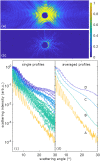
Isolated xenon clusters were irradiated with intense XUV pulses (91 eV photon energy and 3×10¹⁴ W/cm2 peak intensity in the center of the focal spot). A total of 32 events with single clusters of (400±50) nm radius were selected for analysis by the characteristic spacing of the diffraction rings. (a) and (b) Representative diffraction images (second brightest and darkest image of 32 events). (c) Radial profiles of the 32 single-shot images (corrected for the flat detector and nonlinear response, averaged over the scattering angle ϕ; see text). The color coding indicates the binning of events with similar intensities (the least intense category only contains a single pattern). (d) Radial profiles of averaged patterns from bins A to D. For increasing scattered intensity, an upward shift of the profiles (linear response) and an additional modulation of the profiles (corresponding to the ionization and plasma formation) can be observed.
FIG. 2.
FIG. 2.
Absorption of neutral xenon atoms and atomic ions at 91 eV. Total absorption cross sections σabs in Mbarn of neutral Xe, Xe+, Xe2+, Xe3+, Xe4+, and Xe5,6,7+ (colored points). Note that the value of 2 Mbarn for 5 to 7+ constitutes an upper bound. The corresponding penetration depth in nm (black crosses) is calculated using labs=1na·σabs, with na being the atomic density of solid xenon.
FIG. 3.
FIG. 3.
(a) Simulation of the distributions of the relative charge state abundances ρq(x) for a one-dimensional chain of 824 atoms, i.e., 400 nm length. 870 photons (corresponding to 1014W/cm2) fall on the geometric cross section of one xenon atom and are propagated along the chain. Absorption cross sections of atomic xenon and its ions from Fig. 2 are used for calculating absorption probabilities. (b) The average charge state q¯(x) drops from around 6+ to neutral within 80 nm. The relative charge state abundances further allow us to determine an effective absorption index β(x), revealing a transition within 50 nm by an order of magnitude.
FIG. 4.
FIG. 4.
(a) Difference profiles from the averaged profiles shown in Fig. 1(d). For better visibility, the upper curves were shifted by multiplication with a factor. (b) Fitted core–shell Mie profiles using the code from Shen. The refractive index of the core was kept constant to n=1.004+i·0.045 [values of neutral xenon at 91 eV (Ref. 84)]. See text for details. Analog to (a), the profiles II–IV were shifted by a multiplicative factor for better visibility. Dashed lines in (a) and (b) show the profiles below an angle of 10°, where the experimental data were excluded from the fitting process. (c) Parameters of the shell obtained from the fitting, i.e., shell thickness d (in nm), absorption index β, and refractive index decrement δ. (d) Visualization of the sequence of core–shell structures derived from the fitting with changing parameters of the shell [for the exact values of the refractive indices, compare with the 2D color map or with the graphs for β and δ given in (c)].

References

    1. Feldhaus J., Krikunova M., Meyer M., Möller T., Moshammer R., Rudenko A., Tschentscher T., and Ullrich J., “ AMO science at the FLASH and European XFEL free-electron laser facilities,” J. Phys. B 46, 164002 (2013).10.1088/0953-4075/46/16/164002 - DOI
    1. Bostedt C., Boutet S., Fritz D. M., Huang Z., Lee H. J., Lemke H. T., Robert A., Schlotter W. F., Turner J. J., and Williams G. J., Rev. Mod. Phys. 88, 015007 (2016).10.1103/RevModPhys.88.015007 - DOI
    1. Bencivenga F., Capotondi F., Principi E., Kiskinova M., and Masciovecchio C., “ Coherent and transient states studied with extreme ultraviolet and x-ray free electron lasers: Present and future prospects,” Adv. Phys. 63, 327–404 (2014).10.1080/00018732.2014.1029302 - DOI
    1. Yabashi M., Tanaka H., Tanaka T., Tomizawa H., Togashi T., Nagasono M., Ishikawa T., Harries J. R., Hikosaka Y., Hishikawa A., Nagaya K., Saito N., Shigemasa E., Yamanouchi K., and Ueda K., “ Compact XFEL and AMO sciences: SACLA and SCSS,” J. Phys. B 46, 164001 (2013).10.1088/0953-4075/46/16/164001 - DOI
    1. Chapman H. N., Barty A., Bogan M. J., Boutet S., Frank M., Hau-Riege S., Marchesini S., Woods B., Bajt S., Benner W. H., London R. A., Plonjes E., Kuhlmann M., Treusch R., Dusterer S., Tschentscher T., Schneider J. R., Spiller E., Möller T., Bostedt C., Hoener M., Shapiro D. A., Hodgson K. O., van der Spoel D., Burmeister F., Bergh M., Caleman C., Huldt G., Seibert M., Maia F. R. N. C., Lee R. W., Szoke A., Timneanu N., and Hajdu J., “ Femtosecond diffractive imaging with a soft-x-ray free-electron laser,” Nat. Phys. 2, 839–843 (2006).10.1038/nphys461 - DOI