. 2020 May 20;7(5):1096-1103.
doi: 10.1021/acsphotonics.0c00121. Epub 2020 Apr 1.

Monochromatic X-ray Source Based on Scattering from a Magnetic Nanoundulator

Affiliations

Monochromatic X-ray Source Based on Scattering from a Magnetic Nanoundulator

Sophie Fisher et al. ACS Photonics. .

Abstract

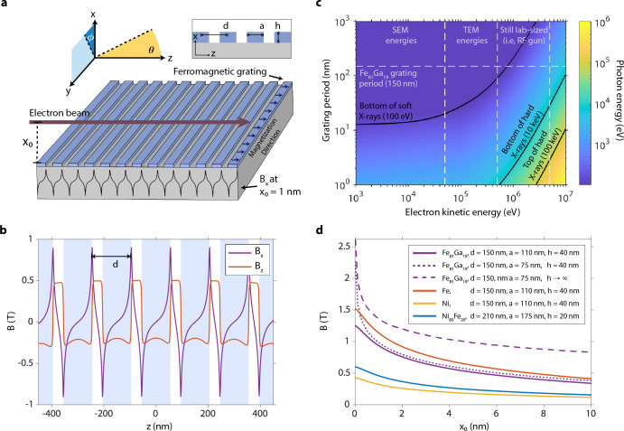
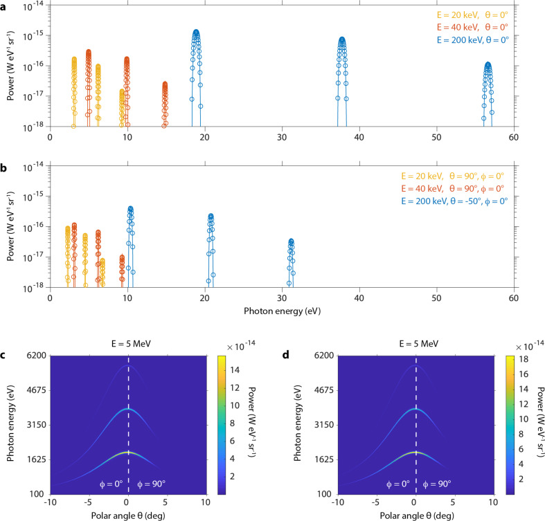
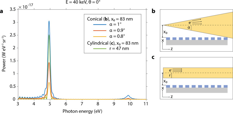
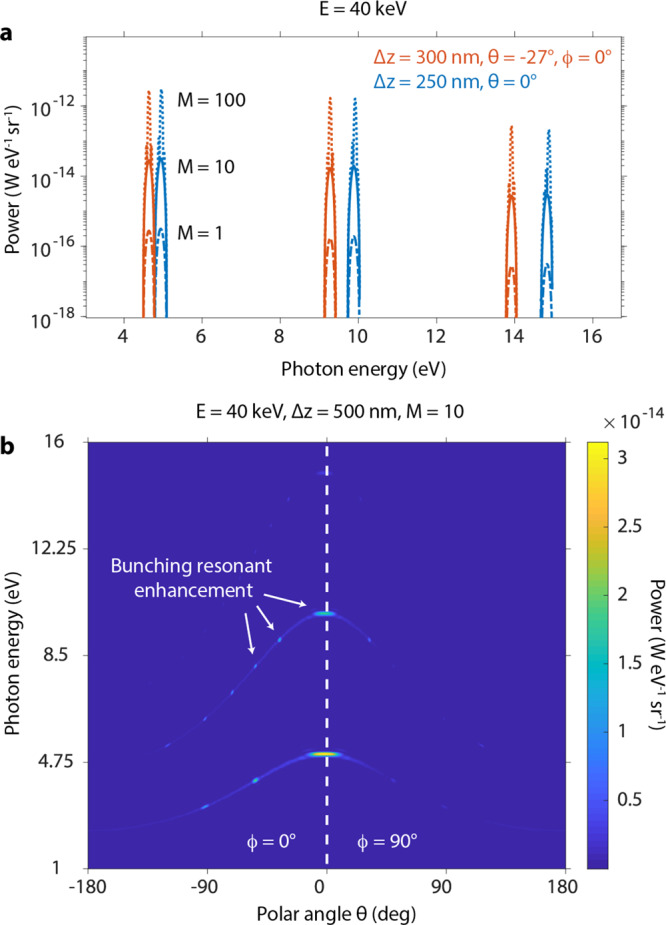
We present a novel design for an ultracompact, passive light source capable of generating ultraviolet and X-ray radiation, based on the interaction of free electrons with the magnetic near-field of a ferromagnet. Our design is motivated by recent advances in the fabrication of nanostructures, which allow the confinement of large magnetic fields at the surface of ferromagnetic nanogratings. Using ab initio simulations and a complementary analytical theory, we show that highly directional, tunable, monochromatic radiation at high frequencies could be produced from relatively low-energy electrons within a tabletop design. The output frequency is tunable in the extreme ultraviolet to hard X-ray range via electron kinetic energies from 1 keV to 5 MeV and nanograting periods from 1 μm to 5 nm. The proposed radiation source can achieve the tunability and monochromaticity of current free-electron-driven sources (free-electron lasers, synchrotrons, and laser-driven undulators), yet with a significantly reduced scale, cost, and complexity. Our design could help realize the next generation of tabletop or on-chip X-ray sources.


Conflict of interest statement

The authors declare no competing financial interest.

Figures

Figure 1
Figure 1
Compact free-electron source of high-frequency radiation. (a) Schematic of the compact radiation source, in which free electrons oscillate in the magnetic near-field of a ferromagnetic nanograting to produce high-frequency radiation. The magnetic field is periodic in z with the nanograting period and has high amplitude (∼1–2 T) toward the nanograting surface (small x0). (b) Plots of the nonzero magnetic field components at a height x0 = 1 nm above the surface, using the parameters of an Fe81Ga19 nanograting fabricated and characterized in ref (16), where μ = 1.4 × 10⁶A/m, d = 150 nm, a = 110 nm, h = 40 nm, and N = 31. z = 0 is defined to be at the center of the nanograting. The positions of the nanograting stripes relative to the field profiles are shown in light blue. (d) The parameters of the nanograting can be tuned to produce a stronger field at the surface by taking the filling ratio a/d to 1/2 (purple, dotted line), increasing the height to period ratio h/d (purple, dashed line), and choosing a ferromagnetic material with a higher magnetization at saturation (orange line). (c) The dispersion relation of the source shows the production of high-frequency radiation ranging from extreme UV to hard X-rays that can be tuned with the nanograting period and electron kinetic energy.
Figure 2
Figure 2
Tunability of the emitted photon energy via the electron energy. (a, b) Numerical (circles) vs analytical (solid lines) results of the radiation power from a single electron in units of power per photon energy per solid angle for a variety of electron energies and fixed polar angles. Different colors represent different electron energies. The polar angles in (b) are chosen such that ϕ = 0 and θ is approximately halfway between the leftmost edge and the peak of the full angular spectrum. Numerical (c) vs analytical (d) results of the full angular spectrum of radiation power from a single electron with energy 5 MeV. All calculations are carried out using the parameters of the reference nanograting with a length along z of 4.65 μm and an electron launched in the z direction with initial height x0 = 1 nm above the nanograting surface.
Figure 3
Figure 3
Dependence of the power spectrum on electron beam geometry. (a) Numerical results of the on-axis radiation power for conical (b) and cylindrical (c) electron beam geometries in units of power per photon energy per solid angle. Conical beams with varying half angles α are considered. The initial height x0 for all geometries is chosen so that the conical beam with the largest half angle (α = 1°) grazes the edge of the nanograting, as in (b). The electron energy is set to 40 keV and the beam brightness to 109 A cm–2 sr–1. All nanograting parameters are as in Figure 2.
Figure 4
Figure 4
Enhanced power spectrum due to a bunched electron beam. (a, b) Radiation power in units of power per photon energy per solid angle due to a bunched electron beam with uniform energy 40 keV launched in the z direction with initial height x0 = 1 nm above the nanograting. The beam is one electron thick and consists of M electrons linearly spaced along z by a distance Δz. (a) The radiation power for varying values of Δz, M, and polar angle θ. The polar angles are chosen so that ϕ = 0° and θ lies on the enhancement occurring closest to θ = 0°. The total radiation power increases by 2 orders of magnitude for every order of magnitude increase in M, the number of bunches. (b) The full angular spectrum of radiation power for a 40 keV electron beam with Δz = 250 nm and M = 10, showing enhancement both on and off axis.

References

    1. Sakdinawat A.; Attwood D. Nanoscale x-ray imaging. Nat. Photonics 2010, 4, 840.10.1038/nphoton.2010.267. - DOI
    1. Hitchcock A. P. Soft X-ray spectromicroscopy and ptychography. J. Electron Spectrosc. Relat. Phenom. 2015, 200, 49–63. 10.1016/j.elspec.2015.05.013. - DOI
    1. Schmitz C.; Wilson D.; Rudolf D.; Wiemann C.; Plucinski L.; Riess S.; Schuck M.; Hardtdegen H.; Schneider C. M.; Tautz F. S.; Juschkin L. Compact extreme ultraviolet source for laboratory-based photoemission spectromicroscopy. Appl. Phys. Lett. 2016, 108, 234101.10.1063/1.4953071. - DOI
    1. Solak H. H. Nanolithography with coherent extreme ultraviolet light. J. Phys. D: Appl. Phys. 2006, 39, R171–R188. 10.1088/0022-3727/39/10/R01. - DOI
    1. McNeil B. W. J.; Thompson N. R. X-ray free-electron lasers. Nat. Photonics 2010, 4, 814.10.1038/nphoton.2010.239. - DOI