. 2020 Jun 17;6(25):eaaz4849.
doi: 10.1126/sciadv.aaz4849. eCollection 2020 Jun.

Guide-free Cas9 from pathogenic Campylobacter jejuni bacteria causes severe damage to DNA

Affiliations

Guide-free Cas9 from pathogenic Campylobacter jejuni bacteria causes severe damage to DNA

Chinmoy Saha et al. Sci Adv. .

Abstract

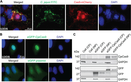
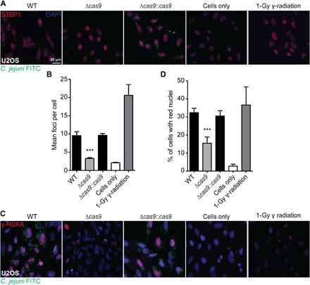
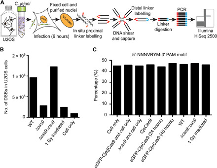
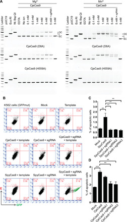
CRISPR-Cas9 systems are enriched in human pathogenic bacteria and have been linked to cytotoxicity by an unknown mechanism. Here, we show that upon infection of human cells, Campylobacter jejuni secretes its Cas9 (CjeCas9) nuclease into their cytoplasm. Next, a native nuclear localization signal enables CjeCas9 nuclear entry, where it catalyzes metal-dependent nonspecific DNA cleavage leading to cell death. Compared to CjeCas9, native Cas9 of Streptococcus pyogenes (SpyCas9) is more suitable for guide-dependent editing. However, in human cells, native SpyCas9 may still cause some DNA damage, most likely because of its ssDNA cleavage activity. This side effect can be completely prevented by saturation of SpyCas9 with an appropriate guide RNA, which is only partially effective for CjeCas9. We conclude that CjeCas9 plays an active role in attacking human cells rather than in viral defense. Moreover, these unique catalytic features may therefore make CjeCas9 less suitable for genome editing applications.


Figures

Fig. 1
Fig. 1. CjeCas9 is released by C. jejuni during infection of human cells and translocate into their nuclei.
(A) Representative microscopic images of human cells infected with C. jejuni bacteria (anti–C. jejuni FITC, green) expressing Cas9-mCherry (red). FITC is fluorescein isothiocyanate, a green fluorescent tracer. (B) Representative microscopic images of nuclear eGFP (enhanced green fluorescent protein)–CjeCas9 localization in human cells (green). The eGFP transfected cells represent a control. (A and B) Nuclei are counterstained with 4′,6-diamidino-2-phenylindole (DAPI) (blue). (C) Nuclear (NP) and cytoplasmic (CP) protein fractions of human cells. Glyceraldehyde phosphate dehydrogenase (GAPDH) verified the quality of the separation.
Fig. 2
Fig. 2. CjeCas9 activates the chromatin and DNA integrity markers, 53BP1 and γ-H2AX, in U2OS cells.
C. jejuni GB11 (WT) and its variants Δcas9 or Δcas9::cas9 were stained with anti–C. jejuni FITC (green), and nuclei were counterstained with DAPI (blue). Gamma (γ)–irradiated cells are used as a positive control, and untreated cells were used as a negative control. (A) Representative microscopic images of human cells infected with WT, Δcas9, or Δcas9::cas9 were stained for 53BP1 (red). (B) The number of 53BP1 related foci per cell (mean foci per cell, y axis) were counted in human cells. The C. jejuni bacteria used are shown on the x axis. (C) Representative microscopic images of human cells infected with WT, Δcas9, or Δcas9::cas9 were stained for γ-H2AX (red). (D) The percentage of human cells with bright red nuclei is displayed on the y axis. The C. jejuni bacteria used are listed at the x axis. Values represent means ± SEM. ***P < 0.001 [one-way analysis of variance (ANOVA), Bonferroni’s multiple comparison test].
Fig. 3
Fig. 3. Analysis of chromosomal DSBs induction using BLESS.
(A) Overview illustration of the BLESS method after C. jejuni infection of U2OS cells. (B) Histogram showing numbers of DSBs accumulated during bacterial infection. Cells that were 1 Gy (gray) irradiated are used as a positive control. Untreated cells were used as a control in the BLESS analyses. (C) Example outcome of PAM analyses. y axis shows the percentage of the 5′-NNNVRYM-3′ PAM motif that is detected in the BLESS-obtained DSBs breaks, as observed in raw and filtered datasets obtained from the samples shown on the x axis.
Fig. 4
Fig. 4. In vitro cleavage assay and in vivo genome editing efficiencies of native CjeCas9 and SpyCas9 in human cells.
(A) CjeCas9 activity and the effect of divalent cations (Mn2+ and Mg2+) concentration on plasmid DNA cleavage. SC is super coiled, L is linear, and OC is open-circle plasmid DNA. (B) Restoration of GFP expression mediated by CjeCas9 or SpyCas9 genome editing in human cells [K562(GFPmut)] and quantified using FACS. In Q1-LR, the percentages of CjeCas9 or SpyCas9 genome edited cells that have a restored GFP expression are visualized (green). Q1-UL are dead cells. Q1-UR are GFP-positive dead cells. Q1-LL are unedited [K562(GFPmut)] cells. (C and D) Percentages of apoptotic human cells (K562) obtained after 6 hours. Concentration of CjeCas9 or SpyCas9 is 30 pmol, and for the sgRNA, the concentrations are 30, 60, and 90 pmol [for single guide RNA1 (sgRNA1), sgRNA2, and sgRNA3, respectively]. Results shown as means ± SEM. ***P < 0.001 and **P < 0.01 (one-way ANOVA, Bonferroni’s multiple comparison test).

References

    1. Mohanraju P., Makarova K. S., Zetsche B., Zhang F., Koonin E. V., van der Oost J., Diverse evolutionary roots and mechanistic variations of the CRISPR-Cas systems. Science 353, aad5147 (2016). - PubMed
    1. Westra E. R., Buckling A., Fineran P. C., CRISPR-Cas systems: Beyond adaptive immunity. Nat. Rev. Microbiol. 12, 317–326 (2014). - PubMed
    1. Louwen R., Staals R. H. J., Endtz H. P., van Baarlen P., van der Oost J., The role of CRISPR-Cas systems in virulence of pathogenic bacteria. Microbiol. Mol. Biol. Rev. 78, 74 (2014). - PMC - PubMed
    1. Haft D. H., Selengut J., Mongodin E. F., Nelson K. E., A guild of 45 CRISPR-associated (Cas) protein families and multiple CRISPR-Cas subtypes exist in prokaryotic genomes. PLoS Comput. Biol. 1, e60 (2005). - PMC - PubMed
    1. Louwen R., Horst-Kreft D., Boer A. G., Graaf L., Knegt G., Hamersma M., Heikema A. P., Timms A. R., Jacobs B. C., Wagenaar J. A., Endtz H. P., Oost J., Wells J. M., Nieuwenhuis E. E. S., Vliet A. H. M., Willemsen P. T. J., Baarlen P., Belkum A., A novel link between Campylobacter jejuni bacteriophage defence, virulence and Guillain–Barré syndrome. Eur. J. Clin. Microbiol. Infect. Dis. 32, 207–226 (2013). - PubMed

Publication types

MeSH terms

LinkOut - more resources