

Three-dimensional-printing for microfluidics or the other way around?
- PMID: 32596534
- PMCID: PMC7294695
- DOI: 10.18063/ijb.v5i2.192
Three-dimensional-printing for microfluidics or the other way around?
Erratum in
-
ERRATUM.Int J Bioprint. 2020 Sep 17;6(4):309. doi: 10.18063/ijb.v6i4.309. eCollection 2020. Int J Bioprint. 2020. PMID: 33102924 Free PMC article.
Abstract
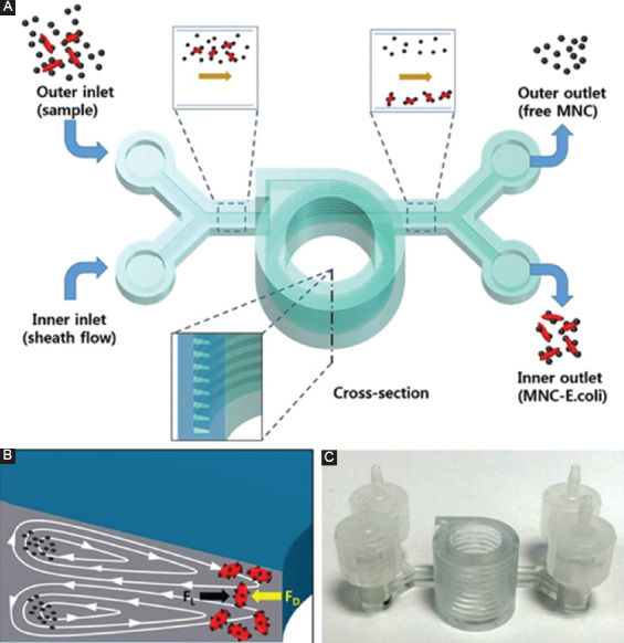
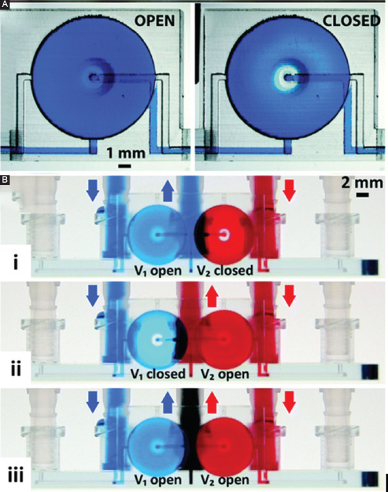
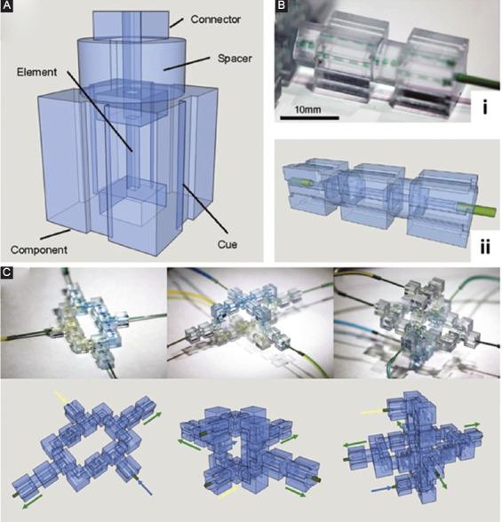
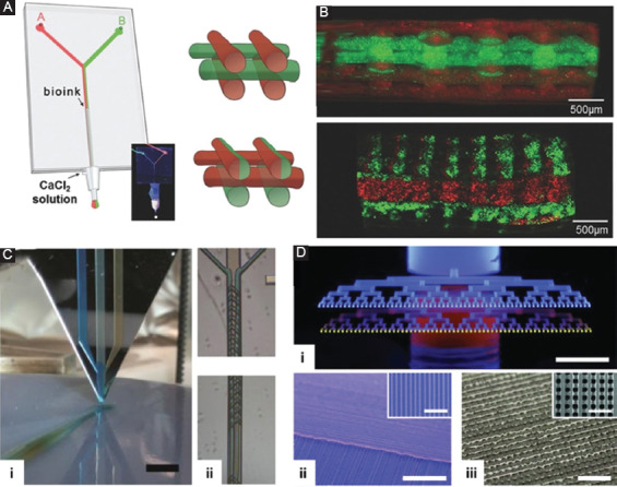
As microfluidic devices are designed to tackle more intricate tasks, the architecture of microfluidic devices becomes more complex, and more sophisticated fabrication techniques are in demand. Therefore, it is sensible to fabricate microfluidic devices by three-dimensional (3D)-printing, which is well-recognized for its unique ability to monolithically fabricate complex structures using a near-net-shape additive manufacturing process. Many 3D-printed microfluidic platforms have been demonstrated but can 3D-printed microfluidics meet the demanding requirements in today's context, and has microfluidics truly benefited from 3D-printing? In contrast to 3D-printed microfluidics, some go the other way around and exploit microfluidics for 3D-printing. Many innovative printing strategies have been made possible with microfluidics-enabled 3D-printing, although the limitations are also largely evident. In this perspective article, we take a look at the current development in 3D-printed microfluidics and microfluidics-enabled 3D printing with a strong focus on the limitations of the two technologies. More importantly, we attempt to identify the innovations required to overcome these limitations and to develop new high-value applications that would make a scientific and social impact in the future.
Keywords: 3D-printing; Bioprinting; Microfluidics.
Copyright: © 2019, Whioce Publishing Pte. Ltd.
Figures
References
-
- Whitesides GM. The Origins and the Future of Microfluidics. Nature. 2006;442(7101):368. - PubMed
-
- Mitchell P. Microfluidics-downsizing Large-scale Biology. Nat Biotechnol. 2001;19(8):717. - PubMed
-
- Yin H, Marshall D. Microfluidics for Single Cell Analysis. Curr Opin Biotechnol. 2012;23(1):110–9. - PubMed
-
- Weibel DB, Whitesides GM. Applications of Microfluidics in Chemical Biology. Curr Opin Chem Biol. 2006;10(6):584–91. - PubMed
-
- Grayson ACR, Shawgo RS, Johnson AM, et al. A bioMEMS Review:MEMS Technology for Physiologically Integrated Devices. Proc IEEE. 2004;92(1):6–21. DOI 10.1109/jproc.2003.820534.
LinkOut - more resources
Full Text Sources
Research Materials