

Application of piezoelectric cells printing on three-dimensional porous bioceramic scaffold for bone regeneration
- PMID: 32596544
- PMCID: PMC7310268
- DOI: 10.18063/ijb.v5i2.1.210
Application of piezoelectric cells printing on three-dimensional porous bioceramic scaffold for bone regeneration
Abstract
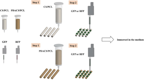
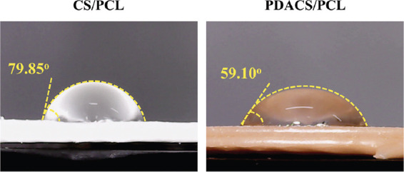
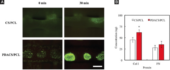
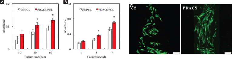
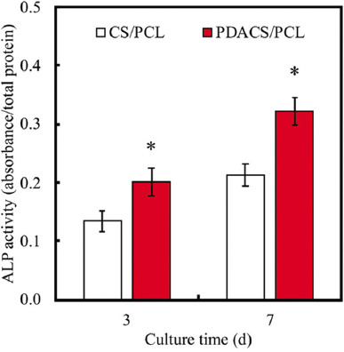
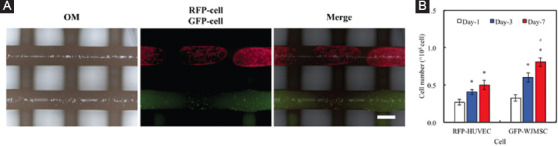
In recent years, the additive manufacture was popularly used in tissue engineering, as the various technologies for this field of research can be used. The most common method is extrusion, which is commonly used in many bioprinting applications, such as skin. In this study, we combined the two printing techniques; first, we use the extrusion technology to form the ceramic scaffold. Then, the stem cells were printed directly on the surface of the ceramic scaffold through a piezoelectric nozzle. We also evaluated the effects of polydopamine (PDA)-coated ceramic scaffolds for cell attachment after printing on the surface of the scaffold. In addition, we used fluorescein isothiocyanate to simulate the cell adhered on the scaffold surface after ejected by a piezoelectric nozzle. Finally, the attachment, growth, and differentiation behaviors of stem cell after printing on calcium silicate/polycaprolactone (CS/PCL) and PDACS/PCL surfaces were also evaluated. The PDACS/PCL scaffold is more hydrophilic than the original CS/PCL scaffold that provided for better cellular adhesion and proliferation. Moreover, the cell printing technology using the piezoelectric nozzle, the different cells can be accurately printed on the surface of the scaffold that provided and analyzed more information of the interaction between different cells on the material. We believe that this method may serve as a useful and effective approach for the regeneration of defective complex hard tissues in deep bone structures.
Keywords: Bone tissue engineering; Calcium silicate; Drop-on-demand; Piezoelectric printing; Polycaprolactone; Polydopamine.
Copyright: © 2019 Shie, et al.
Figures
References
-
- Chiu YC, Fang HY, Hsu TT, et al. 2017, The Characteristics of Mineral Trioxide Aggregate/Polycaprolactone 3-dimensional Scaffold with Osteogenesis Properties for Tissue Regeneration. J Endod. 43:923–9. DOI 10.1016/j.joen.2017.01.009. - PubMed
-
- Sultana A, Ghosh SK, Sencadas V, et al. 2017, Human Skin Interactive Self-powered Wearable Piezoelectric Bio-e-skin by Electrospun Poly-l-lactic Acid Nanofibers for Non-invasive Physiological Signal Monitoring. J Mater Chem B. 5:7352–9. DOI 10.1039/c7tb01439b. - PubMed
-
- Wu Y, Wong YS, Fuh YHJ. 2017, Degradation Behaviors of Geometric Cues and Mechanical Properties in a 3D Scaffold for Tendon Repair. J Biomed Mater Res Part A. 105:1138–49. DOI 10.1002/jbm.a.35966. - PubMed
LinkOut - more resources
Full Text Sources