Review
. 2019 Oct 25;6(2):100-105.
doi: 10.1016/j.wjorl.2019.07.001. eCollection 2020 Jun.

Endoscopic endonasal intraconal orbit surgery

Affiliations
Review

Endoscopic endonasal intraconal orbit surgery

Catherine Banks et al. World J Otorhinolaryngol Head Neck Surg. .

Abstract

Endoscopic endonasal orbital surgery is evolving. With increasing knowledge, expertise, and technology, the historical limits of the endonasal endoscopic approach to the orbit have been redefined. This review discusses the clinical presentation and etiology, and highlights the pertinent anatomy, and discusses the diagnostic workup and surgical approach to orbital tumors and post-operative care. The role of the multidisciplinary team is not to be underestimated. The introduction of a classification system to ensure standardization of technical difficulty and outcome data will assist with international collaboration and further consolidate our attainment of knowledge in this developing field.

Keywords: Endoscopic endonasal orbital surgery; Extraconal space tumors; Intraconal space tumors; Orbital cavernous hemangioma.


Figures

Fig. 1
Fig. 1
Endoscopic approach to the left medial intraconal Space. A: Illustration demonstrates removal of the extraconal fat. The medial and inferior rectus muscles are retracted to highlight the critical anatomy. B: Oculomotor nerve positioned lateral to the medial rectus muscle. The ophthalmic artery enters the orbital apex via the optic canal and is positioned initially lateral to the optic nerve. At approximately 1 cm anterior to the sphenoid face, an inferiomedial muscular trunk (IMT) arises from the ophthalmic artery and sends an arcade of arterioles to supply the medial rectus. C: Arterial branches of the IMT.
Fig. 2
Fig. 2
The anatomical zones of the left orbit. The upper and lower halves of an imaginary line referenced from the medial rectus muscle denote Zone A and Zone B (blue dashed line - not shown in this picture as the medial rectus has been retracted). Zone C is situated posterior to the IMT and represents the most technically challenging, due to the close proximity of the optic nerve and is located approximately 1 cm anterior to the face of the sphenoid.
Fig. 3
Fig. 3
Using three dimensional (3D) reconstruction based on the CT and MRI, the relationship between the tumor and optic nerve can help estimate tumor volume, and assist with spatial confirmation of anatomic lesion relative to other vital structures.
Fig. 4
Fig. 4
Cavernous Hemangioma Exclusively Endonasal Resection (CHEER) staging system. Within the intraconal space, the staging system further subdivides based on the relationship to the IMT of the ophthalmic artery, horizontal axis of the medial rectus, as well as extension to optic canal, inferior orbital fissure, or superior orbital fissure.
Fig. 5
Fig. 5
The location of the orbital lesion within the orbit relative to the optic nerve dictates the choice of the approach. Pathology located with a epicenter medial to the optic nerve or below a “plane of resectability” (POR), which represents a plane subtended by the contralateral nostril and the long axis of the optic nerve, are amenable to the exclusively endoscopic resection (EER). The red shaded area represents the area that is unresectable by the EER.
Fig. 6
Fig. 6
A: Reconstruction of the left medial orbital wall using a right pedicled nasoseptal flap from the contralateral side and positioned over the left extraocular exposed muscles to prevent scarring, reduce delayed contraction and subsequently diplopia and enopthalmos. B: At 3 months post op the nasoseptal flap is well healed and there is a patent sphenoid sinus.

References

    1. Paluzzi A., Gardner P.A., Fernandez-Miranda J.C. Round-the-clock" surgical access to the orbit. J Neurol Surg B Skull Base. 2015;76:12–24. - PMC - PubMed
    1. Shields J.A., Shields C.L., Scartozzi R. Survey of 1264 patients with orbital tumors and simulating lesions: the 2002 Montgomery Lecture, part 1. Ophthalmology. 2004;111:997–1008. - PubMed
    1. Bleier B.S., Castelnuovo P., Battaglia P. Endoscopic endonasal orbital cavernous hemangioma resection: global experience in techniques and outcomes. Int Forum Allergy Rhinol. 2016;6:156–161. - PubMed
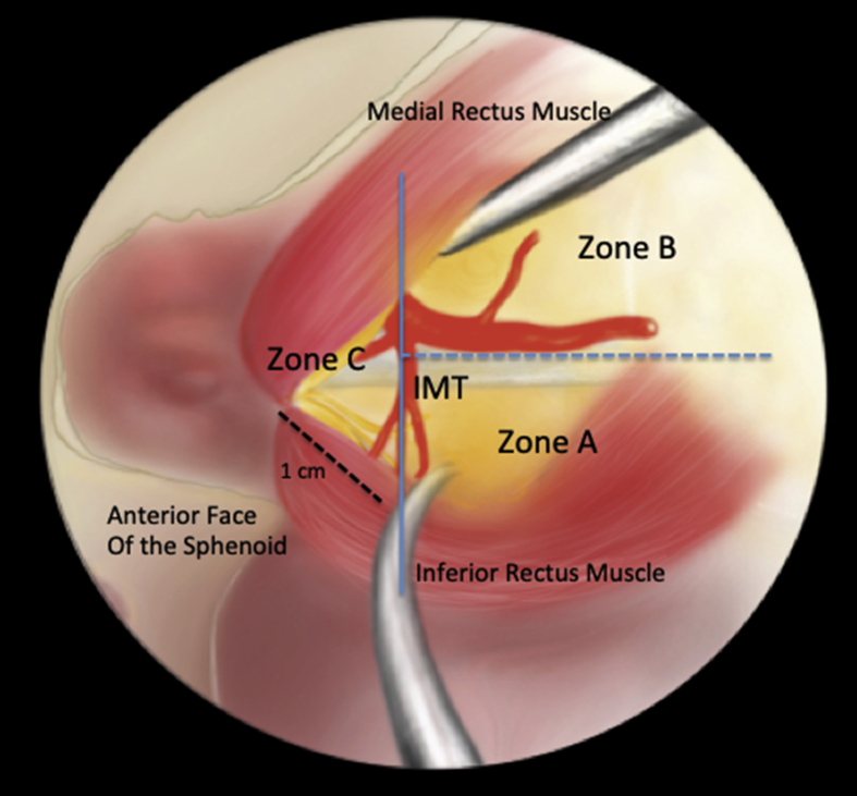
    1. Bleier B.S., Healy D.Y., Chhabra N., Freitag S. Compartmental endoscopic surgical anatomy of the medial intraconal orbital space. Int Forum Allergy Rhinol. 2014;4:587–591. - PubMed
    1. Perrini P., Cardia A., Fraser K., Lanzino G. A microsurgical study of the anatomy and course of the ophthalmic artery and its possibly dangerous anastomoses. J Neurosurg. 2007;106:142–150. - PubMed