. 2020 Aug;24(15):8636-8649.
doi: 10.1111/jcmm.15494. Epub 2020 Jun 28.

HDAC1-mediated deacetylation of LSD1 regulates vascular calcification by promoting autophagy in chronic renal failure

Affiliations

HDAC1-mediated deacetylation of LSD1 regulates vascular calcification by promoting autophagy in chronic renal failure

Jiajun Zhou et al. J Cell Mol Med. 2020 Aug.

Abstract

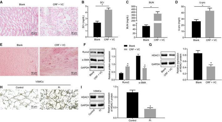
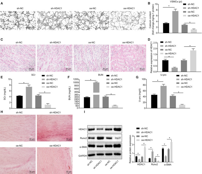
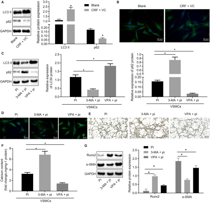
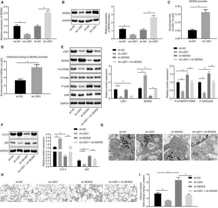
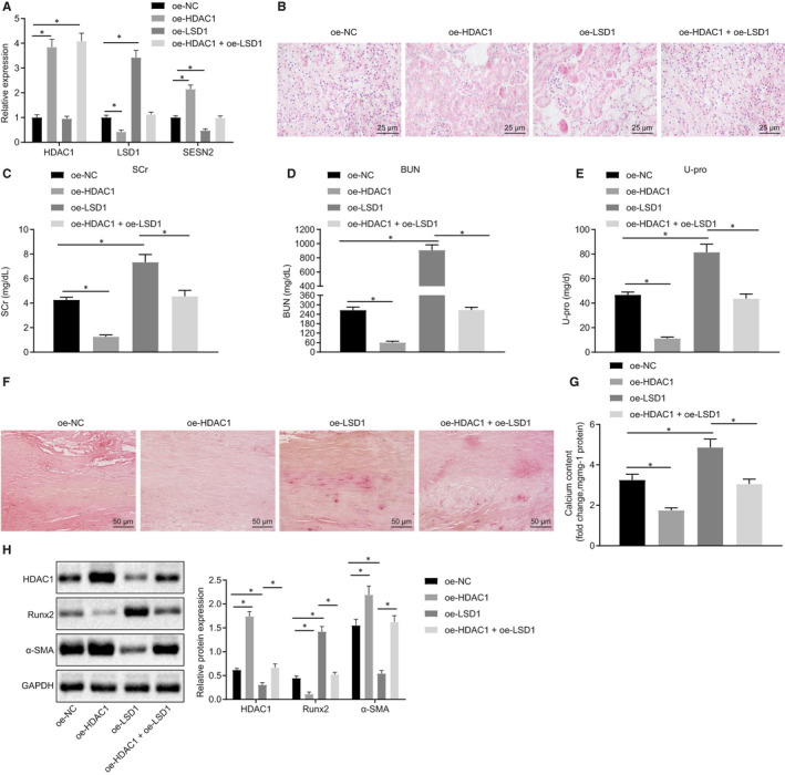
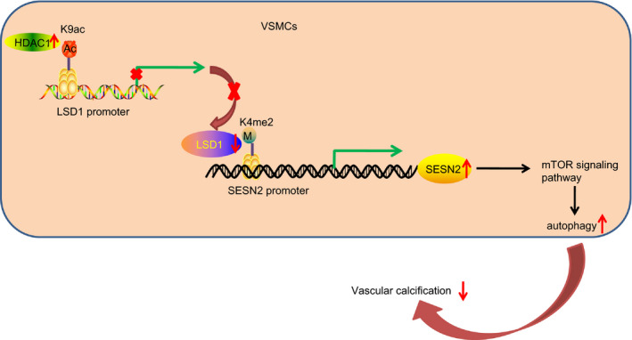
Chronic renal failure (CRF) is commonly associated with various adverse consequences including pathological vascular calcification (VC), which represents a significant clinical concern. Existing literature has suggested the involvement of histone deacetylases (HDACs) in the progression of CRF-induced VC. However, the underlying molecular mechanisms associated with HDACs remain largely unknown. Therefore, we established the adenine-induced CRF rat model and in vitro VC models based on vascular smooth muscle cells (VSMCs) to examine HDAC1/lysine demethylase 1A (LSD1)/SESN2 as a novel molecular pathway in CRF-induced VC. Our initial results demonstrated that HDAC1 reduced the formation of VC in vivo and in vitro. HDAC1 was found to deacetylate LSD1, which subsequently led to impaired transcriptional activity in CRF-induced VC. Moreover, our results illustrated that LSD1 diminished the enrichment of H3K4me2 at the SESN2 promoter. Autophagy was identified as a vasculo-protective element against calcification in VC. Finally, we found that the inhibitory effects of HDAC1 overexpression on VC were partially abolished via over-expressed LSD1 in adenine-induced CRF model rats and in high phosphate-induced VSMCs. Taken together, these results highlight the crucial role of HDAC1 as an antagonistic factor in the progression of VC in CRF, and also revealed a novel regulatory mechanism by which HDAC1 operates. These findings provide significant insight and a fresh perspective into promising novel treatment strategies by up-regulating HDAC1 in CRF.

Keywords: HDAC1; LSD1; SESN2; autophagy; chronic renal failure; deacetylation; vascular calcification.


Conflict of interest statement

The author declares no competing interest exists.

Figures

Figure 1
Figure 1
Down‐regulated HDAC1 is associated with the formation of VC in adenine‐induced CRF rats and high phosphate‐induced VSMCs. A, HE staining of renal tissues (400×). B‐D, Serum levels of SCr, BUN and U‐pro measured by the Falcor 300 analyser. E, Calcification of thoracic aorta determined by Alizarin red staining (200×). F, Protein levels of Runx2 and α‐SMA proteins in aortic tissues determined by Western blot analysis. G, Protein level of HDAC1 protein in aortic tissues determined by Western blot analysis. H, Calcium deposition in VSMCs (200×) determined by Von Kossa staining. I, Protein level of HDAC1 protein in VSMCs induced by high Pi determined by Western blot analysis. BUN, blood urea nitrogen; CRF, chronic renal failure; GAPDH, glyceraldehyde‐3‐phosphate dehydrogenase; HDAC1, histone deacetylase 1; HE, haematoxylin‐eosin; Pi, inorganic phosphate; RT‐qPCR, reverse transcription‐quantitative polymerase chain reaction; Runx2, Runt‐related transcription factor 2; SCr, serum creatinine; U‐pro, urine protein; VC, vascular calcification; VSMCs, vascular smooth muscle cells; α‐SMA, α‐smooth muscle actin. *P < .05 indicates significant difference. Data (mean ± SD) between two groups were analysed using unpaired t test. Data among multiple groups were compared by one‐way analysis of variance (ANOVA) with Tukey's post hoc test. The experiment was performed in triplicate
Figure 2
Figure 2
HDAC1 reduces the formation of VC in adenine‐induced CRF rats and high phosphate‐induced VSMCs. A, Von Kossa staining of calcium deposition in VSMCs (200×). B, Calcium content in cell supernatant measured using a colorimetric method. C, HE staining of renal tissues (400×). D, HDAC1 expression in rats determined using RT‐qPCR. E‐G, Serum levels of SCr, BUN and U‐pro measured by the Falcor 300 analyser. H, Calcification of thoracic aorta (200×) determined by Alizarin red staining. I, Western blot analysis of Runx2 and α‐SMA proteins in aortic tissues. BUN, blood urea nitrogen; CRF, chronic renal failure; GAPDH, glyceraldehyde‐3‐phosphate dehydrogenase; HDAC1, histone deacetylase 1; HE, haematoxylin‐eosin; NC, negative control; oe, overexpression; RT‐qPCR, reverse transcription‐quantitative polymerase chain reaction; Runx2, Runt‐related transcription factor 2; SCr, serum creatinine; sh, short hairpin; U‐pro, urine protein; VC, vascular calcification; VSMCs, vascular smooth muscle cells; α‐SMA, α‐smooth muscle actin. *P < .05 indicates significant difference. Data (mean ± SD) between two groups were analysed using unpaired t test. Data among multiple groups were compared by one‐way analysis of variance (ANOVA) with Tukey's post hoc test. The experiment was conducted in triplicate
Figure 3
Figure 3
Autophagy confers vasculoprotective against calcification in adenine‐induced CRF rats and high phosphate‐induced VSMCs. A, Western blot analysis of LC3 II and p62 proteins in renal tissues. B, Immunofluorescence staining of LC3 II in renal tissues (400×). C, Western blot analysis of LC3 II and p62 proteins in VSMCs. D, Immunofluorescence staining of LC3 II in VSMCs (400×). E, Von Kossa staining of calcium deposition in VSMCs (200×). F, Calcium content in cell supernatant measured using a colorimetric method. G, Western blot analysis of Runx2 and α‐SMA proteins in VSMCs. BUN, blood urea nitrogen; CRF, chronic renal failure; GAPDH, glyceraldehyde‐3‐phosphate dehydrogenase; HDAC1, histone deacetylase 1; Pi, inorganic phosphate; Runx2, Runt‐related transcription factor 2; SCr, serum creatinine; U‐pro, urine protein; VC, vascular calcification; VPA, valproic acid; VSMCs, vascular smooth muscle cells; α‐SMA, α‐smooth muscle actin. *P < .05 indicates significant difference. Data (mean ± SD) between two groups were analysed using unpaired t test, while data among multiple groups were assessed using one‐way ANOVA with Tukey's post hoc test. The experiment was run in triplicate
Figure 4
Figure 4
HDAC1 deacetylates LSD1 and thus mediates autophagy in high phosphate‐induced VC models. A, LSD1 expression in VSMCs without treatment or treated with high Pi detected using RT‐qPCR. B, The enrichment of HDAC1 in the promoter region of LSD1 measured using the ChIP assay. C, The enrichment of H3K9ac in the promoter region of LSD1 measured using ChIP assay. D, LSD1 expression in cells treated with oe‐HDAC1 or SAHA (inhibitor of HDAC1) detected using RT‐qPCR. E, Western blot analysis of LSD1 and HDAC1 protein in cells. F, Western blot analysis of LC3 II and p62 proteins in cells. G, The formation of autophagosomes observed under an electron microscope (20 000×). BUN, blood urea nitrogen; CRF, chronic renal failure; GAPDH, glyceraldehyde‐3‐phosphate dehydrogenase; HDAC1, histone deacetylase 1; HE, haematoxylin‐eosin; NC, negative control; oe, overexpression; Pi, inorganic phosphate; RT‐qPCR, reverse transcription‐quantitative polymerase chain reaction; Runx2, Runt‐related transcription factor 2; SCr, serum creatinine; U‐pro, urine protein; VC, vascular calcification; VPA, valproic acid; VSMCs, vascular smooth muscle cells; α‐SMA, α‐smooth muscle actin. *P < .05 indicates significant difference. Data (mean ± SD) between two groups were analysed using unpaired t test, while data among multiple groups were assessed using one‐way ANOVA with Tukey's post hoc test. The experiment was run in triplicate
Figure 5
Figure 5
LSD1 inhibits autophagy and exacerbates VC by regulation of SESN2 demethylation in high phosphate‐induced VC model mice. A, SESN2 expression in VSMCs detected using RT‐qPCR. B, Western blot analysis of SESN2 protein in VSMCs. C, Enrichment of LSD1 in the promoter region of SESN2 measured by ChIP assay. D, Enrichment of H3K4me2 in the promoter region of SESN2 measured by ChIP assay. E, Western blot analysis of the mTOR signalling pathway‐related proteins. F, Western blot analysis of LC3 II and p62 protein in VSMCs. G, The formation of autophagosomes observed under an electron microscope (20 000×). H, Von Kossa staining of calcium deposition in VSMCs (200×). I, Calcium content in cell supernatant measured by colorimetric method. BUN, blood urea nitrogen; CRF, chronic renal failure; GAPDH, glyceraldehyde‐3‐phosphate dehydrogenase; HDAC1, histone deacetylase 1; HE, haematoxylin‐eosin; NC, negative control; oe, overexpression; RT‐qPCR, reverse transcription‐quantitative polymerase chain reaction; Runx2, Runt‐related transcription factor 2; SCr, serum creatinine; sh, short hair; U‐pro, urine protein; VC, vascular calcification; VPA, valproic acid; VSMCs, vascular smooth muscle cells; α‐SMA, α‐smooth muscle actin. *P < .05 indicates significant difference. Data (mean ± SD) between two groups were analysed using unpaired t test, while data among multiple groups were assessed using one‐way ANOVA with Tukey's post hoc test. The experiment was performed in triplicate
Figure 6
Figure 6
Regulation of the HDAC1/LSD1/SESN2 pathway on VC in adenine‐induced CRF rats. A, The expression of HDAC1, LSD1 and SESN2 in thoracic aorta of rats determined using RT‐qPCR. B, HE staining of pathological changes of renal tissues (400×). C‐E, Serum levels of SCr, BUN, and U‐pro in rats measured by automated analyser Falcor 300. F, Alizarin red staining of calcification of thoracic aorta (200×). G, Calcium content in aortic tissue supernatant measured by colorimetric method. H, Western blot analysis of Runx2 and α‐SMA proteins in aortic tissues. BUN, blood urea nitrogen; CRF, chronic renal failure; GAPDH, glyceraldehyde‐3‐phosphate dehydrogenase; HDAC1, histone deacetylase 1; HE, haematoxylin‐eosin; NC, negative control; oe, overexpression; Pi, inorganic phosphate; RT‐qPCR, reverse transcription‐quantitative polymerase chain reaction; Runx2, Runt‐related transcription factor 2; SCr, serum creatinine; U‐pro, urine protein; VC, vascular calcification; VPA, valproic acid; VSMCs, vascular smooth muscle cells; α‐SMA, α‐smooth muscle actin. *P < .05 indicates significant difference. Data (mean ± SD) between two groups were analysed using unpaired t test. The experiment was run in triplicate
Figure 7
Figure 7
A mechanistic map depicting the potential role of the HDAC1/LSD1/SESN2 pathway in VC in CRF. HDAC1 regulates the deacetylation of LSD1 promoter region through H3K9ac and then inhibits the expression of LSD1. After the inhibition of LSD1, H3K4me2 binds into the promoter region of SESN2 to promote the expression of SESN2. SESN2 promotes autophagy of VSMCs via mTOR signalling pathway regulation which ultimately alleviated VC in CRF. BUN, blood urea nitrogen; CRF, chronic renal failure; HDAC1, histone deacetylase 1; Runx2, Runt‐related transcription factor 2; SCr, serum creatinine; U‐pro, urine protein; VC, vascular calcification; VPA, valproic acid; VSMCs, vascular smooth muscle cells; α‐SMA, α‐smooth muscle actin

Similar articles

Cited by

References

    1. Vadakedath S, Kandi V. Dialysis: a review of the mechanisms underlying complications in the management of chronic renal failure. Cureus. 2017;9:e1603. - PMC - PubMed
    1. Patel MV, Gupta SN, Patel NG. Effects of ayurvedic treatment on 100 patients of chronic renal failure (other than diabetic nephropathy). AYU. 2011;32:483‐486. - PMC - PubMed
    1. Norman JT, Fine LG. Intrarenal oxygenation in chronic renal failure. Clin Exp Pharmacol Physiol. 2006;33:989‐996. - PubMed
    1. Humphris JL, Patch AM, Nones K, et al. Hypermutation in pancreatic cancer. Gastroenterology. 2017;152:68‐74 e2. - PubMed
    1. Lian Y, Xie L, Chen M, Chen L. Effects of an astragalus polysaccharide and rhein combination on apoptosis in rats with chronic renal failure. Evid Based Complement Alternat Med. 2014;2014:271862. - PMC - PubMed

MeSH terms