. 2020 Jun;8(12):e14470.
doi: 10.14814/phy2.14470.

Iron overload causes a mild and transient increase in acute lung injury

Affiliations

Iron overload causes a mild and transient increase in acute lung injury

Vida Zhang et al. Physiol Rep. 2020 Jun.

Abstract

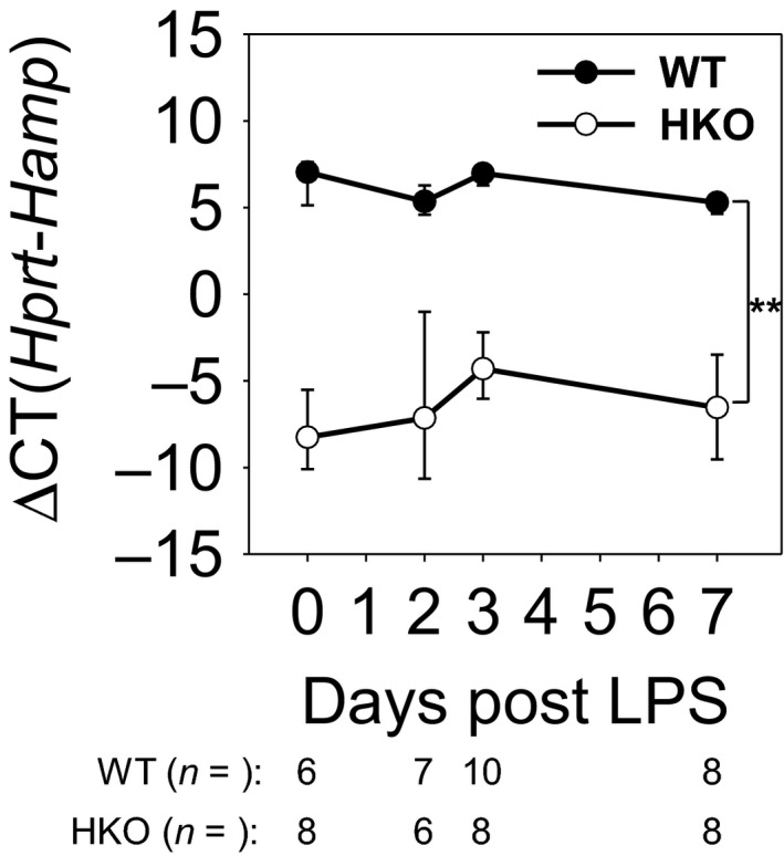
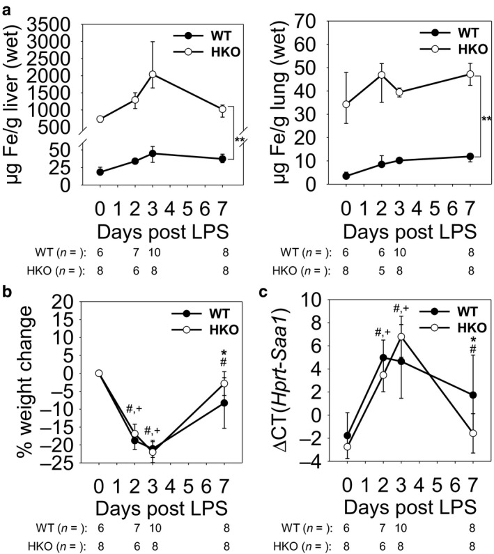
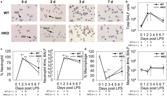
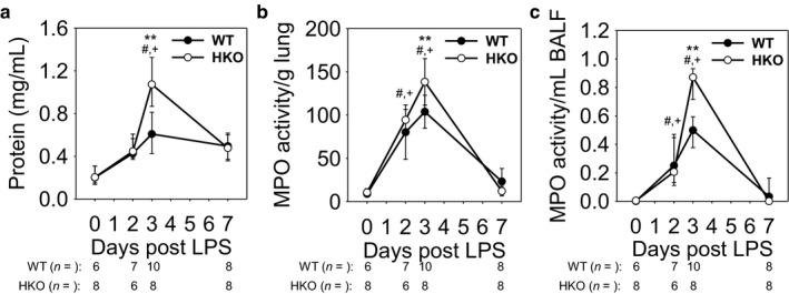
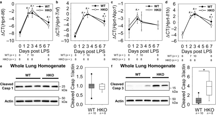
Recent studies have demonstrated a strong link between acute respiratory distress syndrome (ARDS) and the levels of iron and iron-related proteins in the lungs. However, the role of iron overload in ARDS development has yet to be characterized. In this study, we compared the highly iron-overloaded hepcidin knockout mice (HKO) to their iron-sufficient wild-type (WT) littermates in a model of sterile acute lung injury (ALI) induced by treatment with oropharyngeal (OP) LPS. There were no major differences in systemic inflammatory response or airway neutrophil infiltration between the two groups at the time of maximal injury (days 2 and 3) or during the recovery phase (day 7). Hepcidin knockout mice had transiently increased bronchoalveolar lavage fluid (BALF) protein and MPO activity in the lung and BALF on day 3, indicating worse vascular leakage and increased neutrophil activity, respectively. The increased ALI severity in iron-overloaded mice may be a result of increased apoptosis of lung tissue, as evidenced by an increase in cleaved capsase-3 protein in lung homogenates from HKO mice versus WT mice on day 3. Altogether, our data suggest that even severe iron overload has a relatively minor and transient effect in LPS-induced ALI.

Keywords: ARDS; acute lung injury; inflammation; iron overload.


Conflict of interest statement

T.G. and E.N. are shareholders and scientific advisors of Intrinsic LifeSciences and Silarus Therapeutics, and consultants for Ionis Pharmaceuticals, Protagonist, Keryx Pharmaceuticals, La Jolla Pharma, Vifor, Akebia (T.G.), and Gilead (T.G.). Neither A.K. nor V.Z. have any conflicts of interest, financial, or otherwise, to disclose.

Figures

FIGURE 1
FIGURE 1
Hepcidin mRNA levels confirm knockout of hepcidin. WT and HKO mice were treated LPS OP, then analyzed on days 2, 3, and 7. Untreated WT and HKO mice were used as day 0 samples. Liver Hamp mRNA levels confirm knockout of hepcidin. Graph depicts mean ± SD; **p < .001 by a two‐way ANOVA for comparison of genotypes
FIGURE 2
FIGURE 2
Systemic response to LPS aspiration is unaffected by iron overload. WT and HKO mice were treated with LPS OP, then analyzed on days 2, 3, and 7. Untreated WT and HKO mice were used as day 0 samples. (a) Non‐heme iron levels in (left) liver and (right) lung tissues confirm iron overload in HKO mice on high‐iron diet. (b and c) There was no significant difference in whole‐body response to LPS between WT and HKO mice as seen by (b) weight loss or (c) liver Saa1 mRNA levels during peak inflammation. Graphs depict (a) median ± 25th/75th percentile or (b,c) mean ± SD; p < .05 by a two‐way ANOVA for comparison of WT (#) and HKO (+) mice versus day 0, *p < .05 and **p < .001 by a two‐way ANOVA for comparison of genotypes
FIGURE 3
FIGURE 3
LPS induces airway neutrophil infiltration similarly in WT and HKO mice. (a) Representative images of BALF cell cytospins show an evolving leukocyte population. Neut: neutrophil; AM: alveolar macrophages. (b) Increased cell count and (c) % neutrophil and neutrophil count in BALF reflect neutrophil infiltration during the early and recovery phases of inflammation. (d) Though % macrophage in BALF decreases due to influx of neutrophils, absolute macrophage count in BALF remains similar throughout the course of inflammation. Graphs depict (b,c) median ± 25th/75th percentile or (c,d) mean ± SD; p < .05 by a two‐way ANOVA for comparison of WT (#) and HKO (+) mice versus day 0, *p < .05 and **p < .001 by a two‐way ANOVA for comparison of genotypes
FIGURE 4
FIGURE 4
Iron‐overloaded mice develop a mild and transient increase in ALI severity. (a) BALF protein concentration is increased in HKO mice compared to WT mice at 3 d. MPO activity in (b) lung tissue and (c) BALF indicate increased ALI in HKO mice compared to WT mice at 3 d. Graphs depict (a,c) median ± 25th/75thpercentile or (b) mean ± SD; p < .05 by a two‐way ANOVA for comparison of WT (#) and HKO (+) mice versus day 0, **p < .001 by a two‐way ANOVA for comparison of genotypes
FIGURE 5
FIGURE 5
More severe ALI in iron‐overloaded mice may be caused by increased apoptotic cell death. Lung (a) Il6 mRNA and (b) Tnf mRNA show similar induction of lung inflammation between WT and HKO mice. (c) Nqo1 mRNA levels indicate similar ROS generation between WT and HKO mice. Similar lung (d) Il1b mRNA levels and (e) cleaved caspase‐1 levels at 3d between the genotypes indicate that there is no difference in pyroptosis. (f) Cleaved caspase‐3 is increased in HKO mice compared to WT mice at 3 d, suggesting increased apoptosis. Graphs depict (a‐d) mean ± SD or (e, f) median ± 25th/75th percentile; p < .05 by a two way ANOVA for comparison of WT (#) and HKO (+) mice versus day 0, *p < .05 by (b, d) a two‐way ANOVA for comparison of genotypes or (f) Mann Whitney rank‐sum test for comparison of genotypes

Similar articles

Cited by

References

    1. Alber, A. , Howie, S. E. , Wallace, W. A. , & Hirani, N. (2012). The role of macrophages in healing the wounded lung. International Journal of Experimental Pathology, 93, 243–251. 10.1111/j.1365-2613.2012.00833.x - DOI - PMC - PubMed
    1. Bellani, G. , Laffey, J. G. , Pham, T. , Fan, E. , Brochard, L. , Esteban, A. , … Pesenti, A. ; Investigators LS and Group ET . (2016). Epidemiology, patterns of care, and mortality for patients with acute respiratory distress syndrome in intensive care units in 50 countries. JAMA, 315, 788–800. 10.1001/jama.2016.0291 - DOI - PubMed
    1. Benesova, K. , Vujic Spasic, M. , Schaefer, S. M. , Stolte, J. , Baehr‐Ivacevic, T. , Waldow, K. , … Muckenthaler, M. U. (2012). Hfe deficiency impairs pulmonary neutrophil recruitment in response to inflammation. PLoS One, 7, e39363 10.1371/journal.pone.0039363 - DOI - PMC - PubMed
    1. Bice, T. , Cox, C. E. , & Carson, S. S. (2013). Cost and health care utilization in ARDS–different from other critical illness? Seminars in Respiratory and Critical Care Medicine, 34, 529–536. 10.1055/s-0033-1351125 - DOI - PMC - PubMed
    1. Brower, R. G. , Matthay, M. A. , Morris, A. , Schoenfeld, D. , Thompson, B. T. , & Wheeler, A. ; Acute Respiratory Distress Syndrome Network Investigators . (2000). Ventilation with lower tidal volumes as compared with traditional tidal volumes for acute lung injury and the acute respiratory distress syndrome. New England Journal of Medicine, 342, 1301–1308. - PubMed

Publication types