. 2020 Aug;24(15):8779-8788.
doi: 10.1111/jcmm.15513. Epub 2020 Jun 28.

Circ-AKT3 inhibits the accumulation of extracellular matrix of mesangial cells in diabetic nephropathy via modulating miR-296-3p/E-cadherin signals

Affiliations

Circ-AKT3 inhibits the accumulation of extracellular matrix of mesangial cells in diabetic nephropathy via modulating miR-296-3p/E-cadherin signals

Bo Tang et al. J Cell Mol Med. 2020 Aug.

Abstract

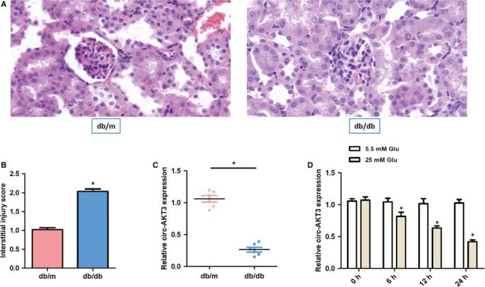
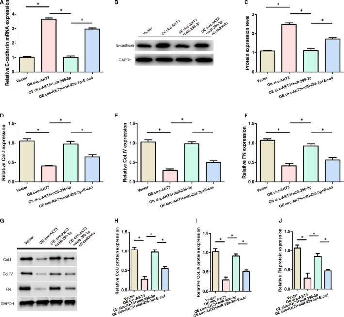
Diabetic nephropathy is a leading cause of end-stage renal disease globally. The vital role of circular RNAs (circRNAs) has been reported in diabetic nephropathy progression, but the molecular mechanism linking diabetic nephropathy to circRNAs remains elusive. In this study, we investigated the significant function of circ-AKT3/miR-296-3p/E-cadherin regulatory network on the extracellular matrix accumulation in mesangial cells in diabetic nephropathy. The expression of circ-AKT3 and fibrosis-associated proteins, including fibronectin, collagen type I and collagen type IV, was assessed via RT-PCR and Western blot analysis in diabetic nephropathy animal model and mouse mesangial SV40-MES13 cells. Luciferase reporter assays were used to investigate interactions among E-cadherin, circ-AKT3 and miR-296-3p in mouse mesangial SV40-MES13 cells. Cell apoptosis was evaluated via flow cytometry. The level of circ-AKT3 was significantly lower in diabetic nephropathy mice model group and mouse mesangial SV40-MES13 cells treated with high-concentration (25 mmol/L) glucose. In addition, circ-AKT3 overexpression inhibited the level of fibrosis-associated protein, such as fibronectin, collagen type I and collagen type IV. Circ-AKT3 overexpression also inhibited the apoptosis of mouse mesangial SV40-MES13 cells treated with high glucose. Luciferase reporter assay and bioinformatics tools identified that circ-AKT3 could act as a sponge of miR-296-3p and E-cadherin was the miR-296-3p direct target. Moreover, circ-AKT3/miR-296-3p/E-cadherin modulated the extracellular matrix of mouse mesangial cells in high-concentration (25 mmol/L) glucose, inhibiting the synthesis of related extracellular matrix protein. In conclusion, circ-AKT3 inhibited the extracellular matrix accumulation in diabetic nephropathy mesangial cells through modulating miR-296-3p/E-cadherin signals, which might offer novel potential opportunities for clinical diagnosis targets and therapeutic biomarkers for diabetic nephropathy.

Keywords: E-cadherin; circ-AKT3; diabetic nephropathy; miR-296-3p.


Conflict of interest statement

The authors confirm that there are no conflicts of interest.

Figures

Figure 1
Figure 1
Circ‐AKT3 might be involved in diabetic nephropathy progression. (A) Representative histological images in two groups. (B) Interstitial injury score in two groups. (C) The relative circ‐AKT3 expression was measured by RT‐PCR in diabetic nephropathy db/db mice compared with matched normal db/m mice (n = 6). (D) The relative expression of circ‐AKT3 was determined by RT‐PCR in mouse mesangial cells (SV40‐MES13) exposed to normal glucose (5.5 mmol/L Glu) and high glucose (25 mmol/L Glu). *P < .05
Figure 2
Figure 2
The overexpression of circ‐AKT3 suppressed the exposed with normal extracellular matrix accumulation of mouse mesangial cells. (A‐C) The overexpression circ‐AKT3 lentivirus plasmid was constructed and its transfection efficiency was confirmed by RT‐PCR. (D‐F) RT‐PCR results indicated the expression levels of Col. I, Col. IV and FN in mouse mesangial cells treated with normal glucose (5.5 mmol/L) or high glucose (25 mmol/L) or transfected with OE circ‐AKT3. (G, H) Relative protein expression of Col. I, Col. IV and FN in mouse mesangial cells treated with normal glucose (5.5 mmol/L) or high glucose (25 mmol/L) or transfected with OE circ‐AKT3. *P < .05. collagen type I (Col. I), collagen type IV (Col. IV) and fibronectin (FN)
Figure 3
Figure 3
Circ‐AKT3 overexpression inhibited the apoptosis of mouse mesangial SV40‐MES13 cells treated with high glucose. (A, B) Cell apoptosis of mouse mesangial SV40‐MES13 cells was detected by flow cytometry assay. (C) Bax, Bcl‐2, Caspase 3 and Cleaved caspase 3 protein expression were measured by Western blot analysis in each group. (D, E) Relative Bax/Bcl‐2 and Cleaved caspase 3/Caspase 3 protein expression were assessed by RT‐PCR in each group. *P < .05
Figure 4
Figure 4
miR‐296‐3p was confirmed to target circ‐AKT3 and E‐cadherin acted as the target protein of miR‐296‐3p. (A) Schematic representation of binding sites between miR‐296‐3p and circ‐AKT3. (B) Relative luciferase activities in mouse mesangial SV40‐MES13 cells cotransfected with the miR‐296‐3p mimic or negative control containing either circ‐AKT3 wild‐type or circ‐AKT3 mutant type. (C) Schematic representation of binding sites between miR‐296‐3p and E‐cadherin mRNA. (D) Relative luciferase activities in mouse mesangial SV40‐MES13 cells cotransfected with the miR‐296‐3p mimic or negative control containing either E‐cadherin mRNA wild‐type or E‐cadherin mRNA mutant type. *P < .05
Figure 5
Figure 5
The correlation between circ‐AKT3, miR‐296‐3p and E‐cadherin. (A) The relative miR‐296‐3p expression was assessed by RT‐PCR in diabetic nephropathy db/db mice compared with matched normal db/m mice (n = 6). (B) The relative E‐cadherin mRNA and protein expression were evaluated by RT‐PCR and Western blot analysis in diabetic nephropathy db/db mice compared with matched normal db/m mice (n = 6). (C) Pearson's correlation analysis of circ‐AKT3 and miR‐296‐3p expressions in db/db diabetic nephropathy mice. (D) Pearson's correlation analysis of circ‐AKT3 and E‐cadherin expressions in db/db diabetic nephropathy mice.*P < .05
Figure 6
Figure 6
circ‐AKT3/miR‐296‐3p/E‐cadherin regulated the extracellular matrix of mouse mesangial cells in high glucose. (A) Relative expression of E‐cadherin mRNA in each group. (B, C) The expression of E‐cadherin protein in each group. (D) Relative Col. I expression in each group. (E) Relative Col. IV expression in each group. (F) Relative FN expression in each group. (G‐J) Relative protein expression of Col. I, Col. IV and FN in each group.*P < .05. collagen type I (Col. I), collagen type IV (Col. IV) and fibronectin (FN)
Figure 7
Figure 7
Effect of circ‐AKT3/miR‐296‐3p/E‐cadherin on the apoptosis of mouse mesangial SV40‐MES13 cells. (A, B) Cell apoptosis of mouse mesangial SV40‐MES13 cells was detected by flow cytometry assay in each group. (C‐E) Relative Bax/Bcl‐2 and Cleaved caspase 3/Caspase 3 protein expression were measured via Western blot analysis in each group *P < .05

References

    1. Magee C, Grieve DJ, Watson CJ, Brazil DP. Diabetic Nephropathy: a Tangled Web to Unweave. Cardiovasc Drugs Ther. 2017;31:579‐592. - PMC - PubMed
    1. Lin YC, Chang YH, Yang SY, Wu KD, Chu TS. Update of pathophysiology and management of diabetic kidney disease. J Formos Med Assoc. 2018;117:662‐675. - PubMed
    1. Flyvbjerg A. The role of the complement system in diabetic nephropathy. Nat Rev Nephrol. 2017;13:311‐318. - PubMed
    1. Ni W‐J, Tang L‐Q, Wei W. Research progress in signalling pathway in diabetic nephropathy. Diabetes Metab Res Rev. 2015;31:221‐233. - PubMed
    1. Chen C, Gong W, Li C, et al. Sphingosine kinase 1 mediates AGEs‐induced fibronectin upregulation in diabetic nephropathy. Oncotarget. 2017;8:78660‐78676. - PMC - PubMed

Publication types