. 2021 Jan 10;34(2):137-153.
doi: 10.1089/ars.2020.8059. Epub 2020 Jul 28.

Mitochondria as Target for Tumor Management of Hemangioendothelioma

Affiliations

Mitochondria as Target for Tumor Management of Hemangioendothelioma

Gayle M Gordillo et al. Antioxid Redox Signal. .

Abstract

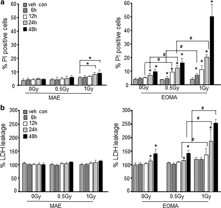
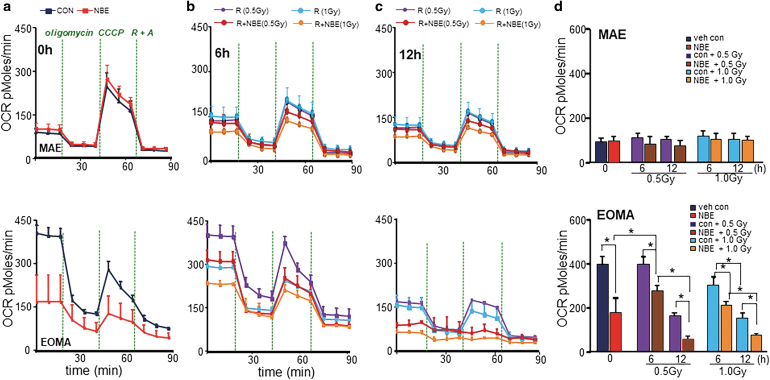
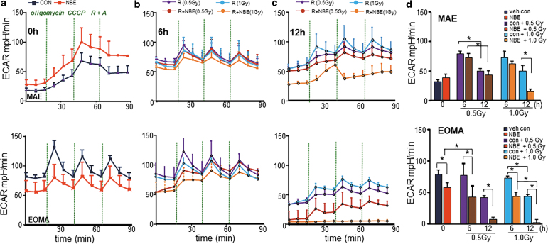
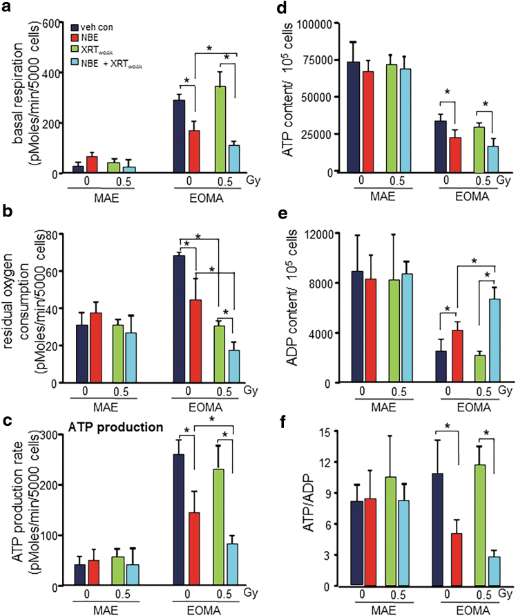
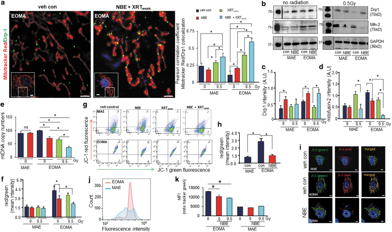
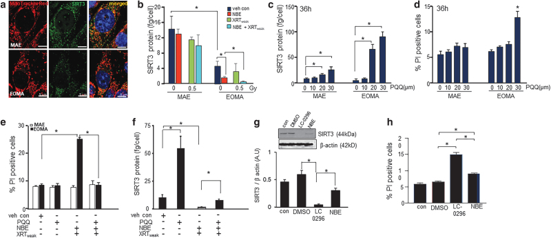
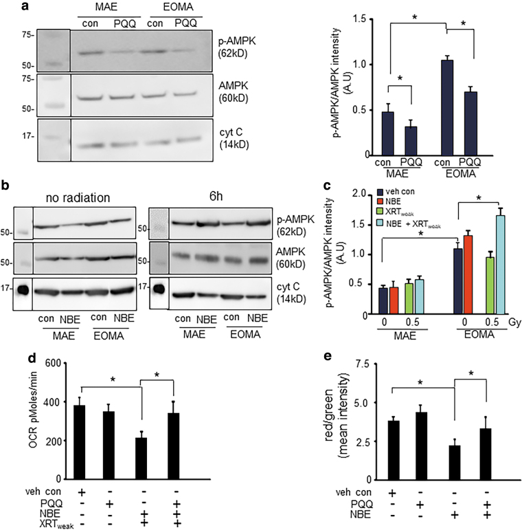
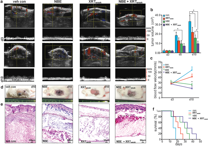
Aims: Hemangioendothelioma (HE) may be benign or malignant. Mouse hemangioendothelioma endothelial (EOMA) cells are validated to study mechanisms in HE. This work demonstrates that EOMA cells heavily rely on mitochondria to thrive. Thus, a combination therapy, including weak X-ray therapy (XRT, 0.5 Gy) and a standardized natural berry extract (NBE) was tested. This NBE is known to be effective in managing experimental HE and has been awarded with the Food and Drug Administration Investigational New Drug (FDA-IND) number 140318 for clinical studies on infantile hemangioma. Results: NBE treatment alone selectively attenuated basal oxygen consumption rate of EOMA cells. NBE specifically sensitized EOMA, but not murine aortic endothelial cells to XRT-dependent attenuation of mitochondrial respiration and adenosine triphosphate (ATP) production. Combination treatment, selectively and potently, influenced mitochondrial dynamics in EOMA cells such that fission was augmented. This was achieved by lowering of mitochondrial sirtuin 3 (SIRT3) causing increased phosphorylation of AMP-activated protein kinase (AMPK). A key role of SIRT3 in loss of EOMA cell viability caused by the combination therapy was evident when pyrroloquinoline quinone, an inducer of SIRT3, pretreatment rescued these cells. Innovation and Conclusion: Mitochondria-targeting NBE significantly extended survival of HE-affected mice. The beneficial effect of NBE in combination with weak X-ray therapy was, however, far more potent with threefold increase in murine survival. The observation that safe natural products may target tumor cell mitochondria and sharply lower radiation dosage required for tumor management warrants clinical testing.

Keywords: hemangioendothelioma; mitochondria; radiation; therapy; tumor.


Conflict of interest statement

The standardized NBE has been commercialized via a university start-up company commercialized as PediaBerry™ in which C.K.S. is a shareholder. All other authors have no competing financial interests.

Figures

FIG. 1.
FIG. 1.
Sensitization of tumor forming endothelial cells to effects of XRT is time and dose dependent. (a) EOMA and MAE cells were treated with NBE (200 μg/mL) and vehicle (1% DMSO) for 6, 12, 24, and 48 h. In some treatment groups, 0.5 and 1 Gy of X-ray radiation was exposed for 6 h after NBE treatment schedule. Cell viability was measured by flow cytometry using PI (10 μg/mL) exclusion, and (b) LDH toxicity assay (MAK066; Sigma-Aldrich) shows a dose-dependent decrease in EOMA cell survival. MAE cells were included as nontumor forming endothelial cell controls. The key indicates the time of sample collection after XRT or after the 24-h pretreatment period for those samples that were not irradiated. *p < 0.05, #p < 0.01, n = 6. DMSO, dimethyl sulfoxide; EOMA, mouse hemangioendothelioma endothelial; LDH, lactate dehydrogenase; MAE, murine aortic endothelial; NBE, natural berry extract; PI, propidium iodide; XRT, X-ray therapy.
FIG. 2.
FIG. 2.
NBE sensitized EOMA, not MAE, cells to weak XRT by inhibiting mitochondrial respiration. NBE/vehicle-treated MAE and EOMA cells were seeded (5000 cells/well) in a Seahorse XF 96-well plate. After the indicated time points of treatment schedule, the OCR was determined during sequential treatments with oligomycin (ATP-synthase inhibitor; 8 mg/mL), CCCP a protonophore that lowers the mitochondrial membrane potential to create conditions for maximal oxidative respiration (100 mM), and antimycin-A (100 mM) + rotenone (100 mM) to inhibit the electron transport chain. (a) Vehicle controls were treated with 1% DMSO. Cells were exposed to XRT alone (0.5/1.0 Gy) or in combination of NBE and XRT for 6-h (b) and 12-h (c) exposure as mentioned earlier. (d) Bar graph comparing basal OCR under all treatment conditions shows decreased OCR after weak XRT exposure in NBE-treated EOMA cells. However, there was no such observation found in the case of MAE cells. Results are expressed as mean ± SD (n = 6). *p < 0.05. ATP, adenosine triphosphate; CCCP, carbonylcyanide-3-chlorophenylhydrazone; OCR, oxygen consumption rate; SD, standard deviation.
FIG. 3.
FIG. 3.
Inhibition of tumor cell glycolysis by NBE alone, and more so in combination with weak XRT. NBE/vehicle-treated MAE and EOMA cells were seeded (5000 cells/well) in a Seahorse XF 96-well plate. After the indicated time points of treatment schedule, the ECAR of cells was determined by a Seahorse XF96 analyzer, upon sequential addition of oligomycin (100 mM), CCCP (100 mM), and antimycin-A (100 mM) + rotenone (100 mM). One of three experiments is shown as representation. (a) Only NBE exposure significantly inhibits ECAR levels in EOMA cells not in MAE. (b) Representative bar graph shows ECAR levels were further inhibited with the exposure of either weak XRT or XRT at 6 h, which was not observed in MAE cells, but with a longer exposure (12 h), MAE cells shows significant reduction in ECAR level (c). (d) The representative bar graph shows decreased ECAR after weak XRT/XRT exposure in NBE-treated EOMA cells. However, only NBE with XRT exposure in MAE cell causes inhibition of ECAR activity. Thus, the 0.5 Gy dose was selected for all our in vitro doses to verify the effects. Results are expressed as mean ± SD (n = 6). *p < 0.05. ECAR, extracellular acidification rate.
FIG. 4.
FIG. 4.
Blunted cellular respiration and reduced ATP production in EOMA cells. (a) Basal respiration measured from Seahorse XF96 was significantly inhibited with weak XRT in NBE-treated EOMA compared with MAE. (b) The residual oxygen consumption also gets compromised with weak XRT exposure in NBE-treated EOMA cells. No such effects were observed in nontumor forming MAE cells. (c) Weak XRT exposure causes a decreased rate of ATP production in NBE-treated EOMA cells compared with the no weak XRT group. All calculations were made using the Agilent Seahorse software provided with the instrument. Results are expressed as mean ± SD (n = 6). (d) Basal cellular ATP, ADP (e), and the ratio (f) were measured in different treatment groups using the EnzyLightTM ADP/ATP ratio assay kit. Results are expressed as mean ± SD (n = 4). *p < 0.05.
FIG. 5.
FIG. 5.
Mitochondrial fission and membrane potential. (a) Colocalization analysis of mitochondrial fission marker (Drp1, ab184247 1:200; Abcam, Cambridge, MA) and MitoTracker Red CMXRos (250 nM) in MAE and EOMA cells before and after treatment with NBE or XRT (n = 3–5). Scale bar = 2 μm. Inset shows the cell from which the region of interest was taken. Scale bar = 5 μm. Complete image set is presented as Supplementary Figure S2. (b) Western blot images of Mfn-2 and Drp1 protein expressions were compared between no-XRT group and weak XRT exposure in NBE- or vehicle-treated MAE/EOMA cells. (c) Bar graph for Drp1 and (d) Mfn-2 protein expressions, normalized with β-actin. (e) Mitochondrial copy numbers of MAE and EOMA cells exposed to vehicle control or NBE. In addition, mitochondrial copy number determination was done for EOMA cells further exposed to weak XRT with or without NBE (n = 3). (f) Loss of mitochondrial membrane potential in EOMA and MAE cells exposed to weak XRT with or without NBE, as assessed by JC-1 flow cytometry. Cells were collected 6 h after XRT exposure with or without NBE treatment and stained with 2.5 μM of JC-1 for 15 min. The ratio of red to green shows a significant decrease of mitochondrial membrane potential in combined treatment of NBE and XRT than XRT alone. Results are expressed as mean ± SD (n = 6). (g) Representative images of cell populations analyzed by the flow cytometer (Beckman-Coulter), using a 488 laser, following standard protocols provided by JC-1 manufacturers. (h) Intensity calculation and (i) representative airyscan confocal (ZEISS LSM 880) images of cells subjected to JC-1 staining (2.5 μM, 30 min) with or without NBE treatment in EOMA cells compared with untreated MAE cells. (j) Flow cytometric profiles of mitochondrial mass levels in EOMA and MAE cells after exposed to NBE, by MitoTracker green FM (25 nM). (k) MFI of MitoTracker green FM of EOMA and MAE cells exposed to weak XRT with or without NBE. Results are expressed in mean ± SE (n = 6). *p < 0.05. Drp1, dynamin-related protein 1; MFI, median fluorescence intensity; Mfn-2, mitofusin 2; SE, standard error.
FIG. 6.
FIG. 6.
SIRT3 depletion responsible for inducible death of EOMA. (a) Representative image of intracellular SIRT3 localization in MAE and EOMA cells. (b) EOMA and MAE cells were seeded in equal numbers (100,000 cells/well) in a 12-well plate. After the mentioned treatment schedule, cells were isolated and measured for SIRT3 level using the SIRT3 ELISA kit. SIRT3 levels was normalized to the number of cells seeded per well. Sirt3 levels were significantly reduced in the cellular supernatant of EOMA compared with MAE, and further reduced when treated with NBE alone or a combined exposure of NBE and weak XRT in EOMA cells. (c) PQQ, a chemical activator of Sirt 3, causes induction of SIRT3 in a dose-dependent manner in both MAE and EOMA. (d) Significant cell death was observed in EOMA cells with 30 μM as shown by PI positivity. (e) Twelve hours pretreatment with PQQ (20 μM) significantly rescued the cellular survival of EOMA cells compared with combined exposure of NBE and weak XRT, and subsequently, the level of Sirt3 was found to be significantly elevated (f) at that point. Results are expressed as mean ± SD (n = 3). (g) Western blot images of SIRT3 protein expression were compared between control-, vehicle control (DMSO)-, LC-0296 (10 μM)-, and NBE-treated groups in EOMA cells. Signal intensities of SIRT3 protein are normalized with β-actin. Results are expressed as mean ± SD (n = 3). (h) Percentage of PI fluorescence expressing cells was counted by flow cytometry, after staining NBE- and LC-0296-treated EOMA cells with PI (10 μg/mL), using the standard protocol. Data are expressed as mean ± SEM (n = 6). *p < 0.05. ELISA, enzyme-linked immunosorbent assay; PQQ, pyrroloquinoline quinone; SEM, standard error of the mean; SIRT3, sirtuin 3.
FIG. 7.
FIG. 7.
AMPK phosphorylation caused by severe depletion of SIRT3 compromises mitochondrial respiration in EOMA cells. (a) Western blot shows elevated ratio of phospho-AMPK to AMPK in EOMA cells compared with MAE. PQQ treatment reduces the AMPK activation by inhibiting phosphorylation in both MAE and EOMA cells. (b) Western blot images of phospho-AMPK and AMPK in NBE alone or combined exposure of NBE and weak XRT. (c) Intensity calculation confirms that either NBE alone or in combination with weak XRT increases the phosphorylation of AMPK in EOMA cells. Results are expressed as mean ± SD (n = 3). (d) OCR data show that pretreatment with PQQ rescued the combined effect of NBE and weak XRT. (e) Intensity calculation of EOMA cells subjected to JC-1 staining with or without NBE or PQQ treatment. Results are expressed as mean ± SD (n = 7). *p < 0.05. AMPK, AMP-activated protein kinase.
FIG. 8.
FIG. 8.
Improved survival of mice with HE tumor. Syngeneic 6- to 8–week-old female 129 P/3 mice received a subcutaneous injection of EOMA cells. Mice were treated with NBE oral gavage (20 mg/kg) and topical application (200 mg/kg) once daily. XRT exposure using irradiator RS2000 was carried out with 2.5 Gy gamma XRT per dose directed at the tumor given on the 3rd, 5th, and 7th day after EOMA cell injection for a total dose of 7.5 Gy. (a) Ultrasound imaging of animals from each group (n = 5) was performed on a Vevo 2100 system (Visual-Sonics, Toronto, Canada) using high-frequency linear array transducers operating between 8 and 17 MHz. (b) Tumor volume was quantified using calipers (length × width × height). Significantly reduced tumor growth was observed at day 10 in NBE+XRT-treated group compared with others. (c) Tumor blood flow analyzed using the Vevo 2100 system shows a significant difference at day 10 after NBE+XRT combined treatment compared with other groups. (d) Representative images of tumor in different treatment groups. (e) H&E staining of tumor section after 10 days of cell injection shows decreased cellular granularity. (f) Kaplan–Meier survival curve analyzed by log-rank analysis shows mice treated with vehicle, XRT only, NBE only, and NBE+XRT. Results are expressed as mean ± SD (n = 5). Here, NBE-only and NBE+XRT treatment has a significant survival advantage compared with vehicle control, *p < 0.05. Also, NBE+XRT treatment has a significant survival advantage compared with XRT only, *p < 0.05. HE, hemangioendothelioma; H&E, hematoxylin and eosin.

References

    1. Alhazzazi TY, Kamarajan P, Xu Y, Ai T, Chen L, Verdin E, and Kapila YL. A novel sirtuin-3 inhibitor, LC-0296, inhibits cell survival and proliferation, and promotes apoptosis of head and neck cancer cells. Anticancer Res 36: 49–60, 2016 - PMC - PubMed
    1. Aminzadeh MA, Rogers RG, Fournier M, Tobin RE, Guan X, Childers MK, Andres AM, Taylor DJ, Ibrahim A, Ding X, Torrente A, Goldhaber JM, Lewis M, Gottlieb RA, Victor RA, and Marban E. Exosome-mediated benefits of cell therapy in mouse and human models of Duchenne muscular dystrophy. Stem Cell Reports 10: 942–955, 2018 - PMC - PubMed
    1. Ansari A, Rahman MS, Saha SK, Saikot FK, Deep A, and Kim KH. Function of the SIRT3 mitochondrial deacetylase in cellular physiology, cancer, and neurodegenerative disease. Aging Cell 16: 4–16, 2017 - PMC - PubMed
    1. Atalay M, Gordillo G, Roy S, Rovin B, Bagchi D, Bagchi M, and Sen CK. Anti-angiogenic property of edible berry in a model of hemangioma. FEBS Lett 544: 252–257, 2003 - PubMed
    1. Aune D, Giovannucci E, Boffetta P, Fadnes LT, Keum N, Norat T, Greenwood DC, Riboli E, Vatten LJ, and Tonstad S. Fruit and vegetable intake and the risk of cardiovascular disease, total cancer and all-cause mortality-a systematic review and dose-response meta-analysis of prospective studies. Int J Epidemiol 46: 1029–1056, 2017 - PMC - PubMed

Publication types

LinkOut - more resources