. 2020 Aug;9(16):e2000536.
doi: 10.1002/adhm.202000536. Epub 2020 Jun 29.

Heparin-Coated Albumin Nanoparticles for Drug Combination in Targeting Inflamed Intestine

Affiliations

Heparin-Coated Albumin Nanoparticles for Drug Combination in Targeting Inflamed Intestine

Sufeng Zhang et al. Adv Healthc Mater. 2020 Aug.

Abstract

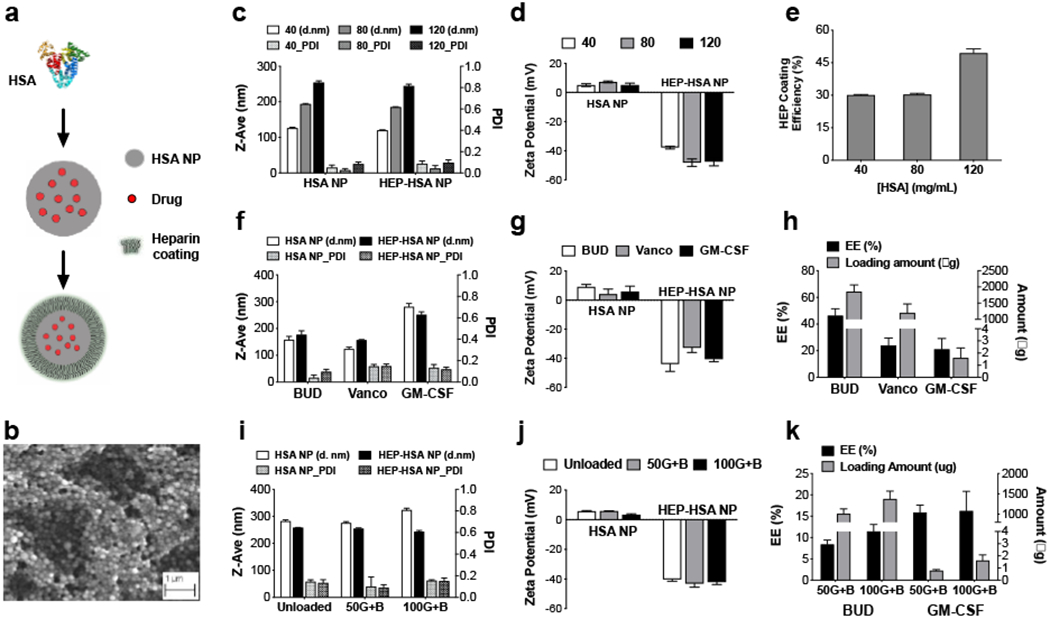
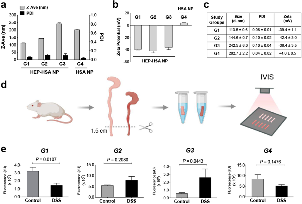
Targeting areas of inflammation offers potential therapeutic and diagnostic benefits by maximizing drug and imaging marker on-target effects while minimizing systemic exposure that can be associated with adverse side effects. This strategy is particularly beneficial in the management of inflammatory bowel disease (IBD). Here an inflammation-targeting (IT) approach based on heparin-coated human serum albumin nanoparticles (HEP-HSA NPs) that utilize the increased intestinal permeability and changes in electrostatic interaction at the site of intestinal inflammation is described. Using small-molecule and biologic drugs as a model for drug combination, the HEP-HSA NPs demonstrate the capacity to load both drugs simultaneously; the dual-drug loaded HEP-HSA NPs exhibit a higher anti-inflammatory effect than both of the single-drug loaded NPs in vitro and selectively bind to inflamed intestine after enema administration in vivo in a murine model of colitis. Importantly, analyses of the physicochemical characteristics and targeting capacities of these NPs indicate that HEP coating modulates NP binding to the inflamed intestine, providing a foundation for future IT-NP formulation development.

Keywords: drug combination; drug delivery; intestinal inflammation; nanoparticles.


Conflict of interest statement

Conflict of Interest

S.Z., J.R.K. and G.T. are co-inventors on a provisional patent application encompassing the technology described in this manuscript.

Figures

Figure 1.
Figure 1.. Synthesis and characterization of heparin-coated human serum albumin nanoparticles (HEP-HSA NPs).
(a) Schematic outlining the formation of drug-loaded HEP-HSA NPs. (b) A scanning electron microscopic image of HEP-HSA NPs. (c, d) Size, polydispersity index (PDI), and zeta potential measurement of NPs prepared from 40, 80, and 120 mg mL−1 of HSA, before and after HEP coating. (e) Coating efficiency (%) of 1.0 mg mL−1 HEP in water on different HSA NPs. (f, g) Size, PDI, and zeta potential of HSA NPs loaded with different drugs, before and after HEP coating. BUD: budesonide; Vanco: vancomycin; GM-CSF: granulocyte macrophage colony-stimulating factor. (h) Encapsulation efficiency (EE) and the amount of drug loaded in HSA NPs. (i, j) Size, PDI, and zeta potential of unloaded and dual-drug loaded HSA NPs with BUD (3 mg mL−1) and GM-CSF (50 and 100 μg mL−1, respectively), before and after HEP coating. (k) EE and the amount of BUD and GM-CSF in the dual-drug loaded HSA NPs. Data are presented as mean ± SD from a representative experiment (n = 3; independent experiment was performed at least twice).
Figure 2.
Figure 2.. Dual-drug loaded HEP-HSA NPs demonstrate higher anti-inflammatory effect than single-drug loaded HEP-HSA NPs evaluated in vitro.
(a, b) Size, PDI, and zeta potential measurement of unloaded (UNP), single-drug loaded (B/NP, G/NP), and dual-drug loaded (G+B/NP) HSA NPs, before and after HEP coating. (c, d) EE and the amount of BUD (c) and GM-CSF (d) in single-drug loaded NPs (B/NP, G/NP) and dual-drug loaded NPs (G+B/NP). (e) Comparison of different NP formulations on the inhibition of TNF-α expression by RAW264.7 macrophages. NC: Untreated, non-activated macrophages as negative control; PC: LPS-activated macrophages without NP treatment as positive control. * p = 0.0472 for B/HNP with PC, ** p = 0.0012 for G+B/NP with PC, and p = 0.4775 for G/NP and UNP with PC (Holm-Sidak’s multiple comparison test). (f) Comparison of different NP formulations on the inhibition of NO expression by RAW264.7 macrophages. All NP formulations significantly reduced NO expression compared with PC. **** p < 0.0001 for G+B/NP compared with PC (Holm-Sidak’s multiple comparison test). Data are presented as mean ± SD from a representative experiment (n = 3; independent experiment was performed at least twice).
Figure 3.
Figure 3.. Larger HEP-HSA NPs, not smaller or uncoated ones, exhibited preferential binding to the inflamed colon evaluated ex vivo.
(a - c) Size, PDI, and zeta potential of different Alexa Fluor®680-labeled NP formulations (G1, G2, G3, and G4) for the ex vivo evaluation. (d) Schematic outlining the experimental workflow. The distal colons of mice with dextran sulfate sodium (DSS)-induced colitis and healthy controls were incubated with NPs under 37°C for 30 min, washed, and imaged by IVIS. (e) IVIS fluorescence intensity of G1, G2, G3, and G4 NPs evaluated with mouse colons ex vivo. Control: colons from healthy mice; DSS: colons from mice with DSS-induced colitis. The total fluorescence intensity was determined in a standard-sized region of interest (ROI) around the individual colon pieces with background fluorescence subtracted. Data are presented as mean ± SD (n = 3) in (a - c) and presented as mean ± SEM (n = 3 - 5 mice per group) in (e); P-values were determined by Student’s t test.
Figure 4.
Figure 4.. Larger HEP-HSA NPs loaded with drug combinations displayed preferential binding to the inflamed colon in mice in vivo.
(a, b) Size, PDI, and zeta potential measurement of dual-drug loaded, Alexa Fluor®680-labeled HSA NPs, before and after HEP coating. (c) Schematic of colitis induction in mice and treatment regimen. (d) IVIS imaging of mouse colons dissected from healthy controls (Control) and mice with DSS-induced colitis (DSS) 3h after the enema treatment. (e) IVIS quantification of the fluorescence intensity in (d). The total fluorescence intensity was determined in a standard-sized ROI around the individual colon pieces with background fluorescence subtracted. Data are presented as mean ± SD (n = 3) in (a - b) and presented as mean ± SEM (n = 7 mice per group) in (e); P-values were determined by Student’s t test.
Figure 5.
Figure 5.. Smaller HEP-HSA NPs and uncoated HSA NPs did not exhibit preferential binding to the inflamed colon in mice in vivo.
(a, b) Size, PDI, and zeta potential measurement of Alexa Fluor®680-labeled (a) HEP-HSA NPs prepared from 40 mg mL−1 HSA and (b) uncoated HSA NPs prepared from 120 mg mL−1 HSA for in vivo evaluation, respectively. The treatment regimen was the same as in Figure 4c. The 3-cm distal colons from healthy mice (Control) and colitic mice (DSS) were excised for IVIS imaging (middle) and quantification (right) for (a) and (b), respectively. (c, d) Individual mouse colons after IVIS imaging were divided equally into three 1-cm pieces for fluorescence quantification (left) and myeloperoxidase (MPO) activity (middle) after homogenization, as well as for histology assessment (right) for (a) and (b), respectively. Data are presented as mean ± SD (n = 3) in (a - b) and all data on fluorescence intensity, MPO activity, and histology scores are presented as mean ± SEM (n = 6 - 7 mice per group) in (a - d); P-values were determined by Student’s t test.

References

    1. Kaser A, Zeissig S, Blumberg RS, Annu Rev Immunol 2010, 28, 573–621. - PMC - PubMed
    1. Cho JH, Nat Rev Immunol 2008, 8, 458–466; - PubMed
    2. Chassaing B, Darfeuille-Michaud A, Gastroenterology 2011, 140, 1720–1728. - PubMed
    1. Lichtenstein GR, Loftus EV, Isaacs KL, Regueiro MD, Gerson LB, Sands BE, Am J Gastroenterol 2018, 113, 481–517; - PubMed
    2. Rubin DT, Ananthakrishnan AN, Siegel CA, Sauer BG, Long MD, Am J Gastroenterol 2019, 114, 384–413. - PubMed
    1. Her M, Kavanaugh A, J Allergy Clin Immunol 2016, 137, 19–27; - PubMed
    2. Stallmach A, Hagel S, Bruns T, Best Pract Res Clin Gastroenterol 2010, 24, 167–182. - PubMed
    1. Hoivik ML, Moum B, Solberg IC, Cvancarova M, Hoie O, Vatn MH, Bernklev T, Group IS, Inflamm Bowel Dis 2012, 18, 1540–1549; - PubMed
    2. Ng SC, Shi HY, Hamidi N, Underwood FE, Tang W, Benchimol EI, Panaccione R, Ghosh S, Wu JCY, Chan FKL, Sung JJY, Kaplan GG, Lancet 2018, 390, 2769–2778. - PubMed

Publication types