. 2020 Aug 3;130(8):4218-4234.
doi: 10.1172/JCI138103.

Epithelial-derived gasdermin D mediates nonlytic IL-1β release during experimental colitis

Affiliations

Epithelial-derived gasdermin D mediates nonlytic IL-1β release during experimental colitis

Katarzyna Bulek et al. J Clin Invest. .

Abstract

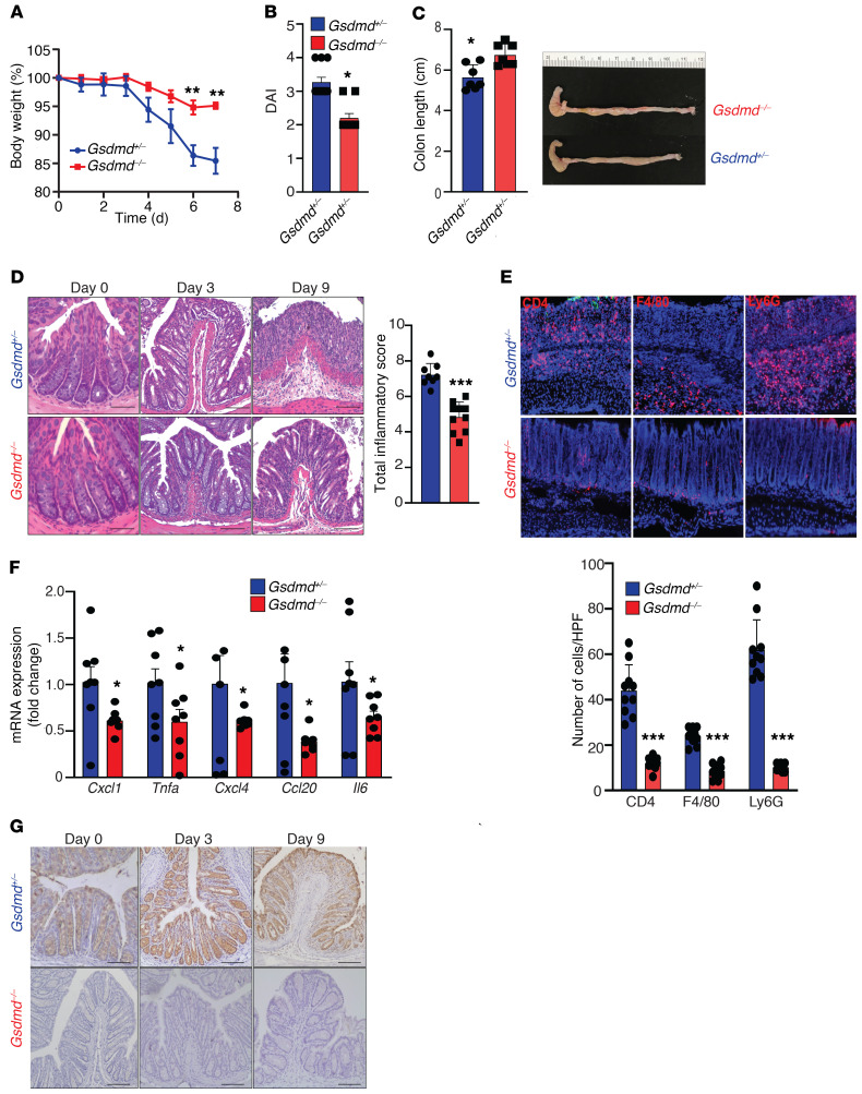
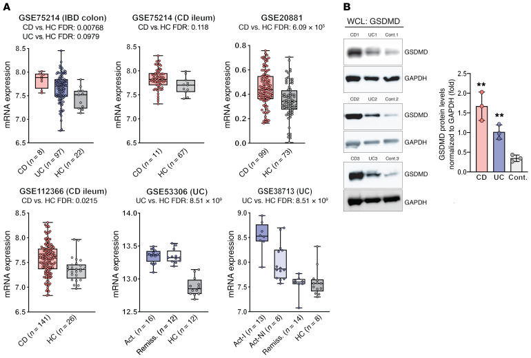
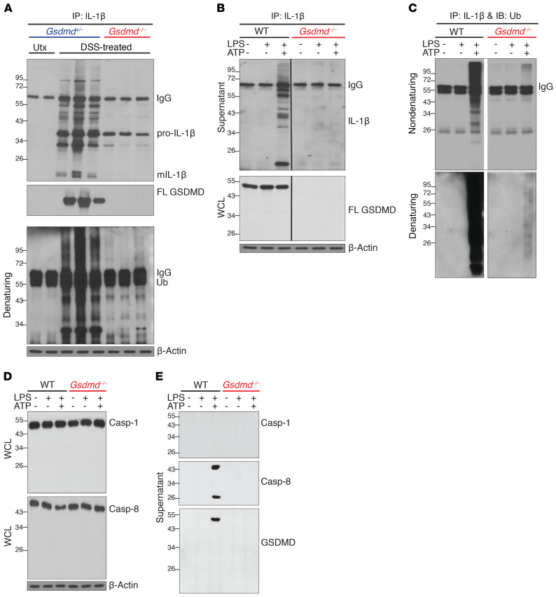
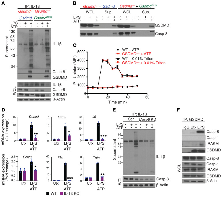
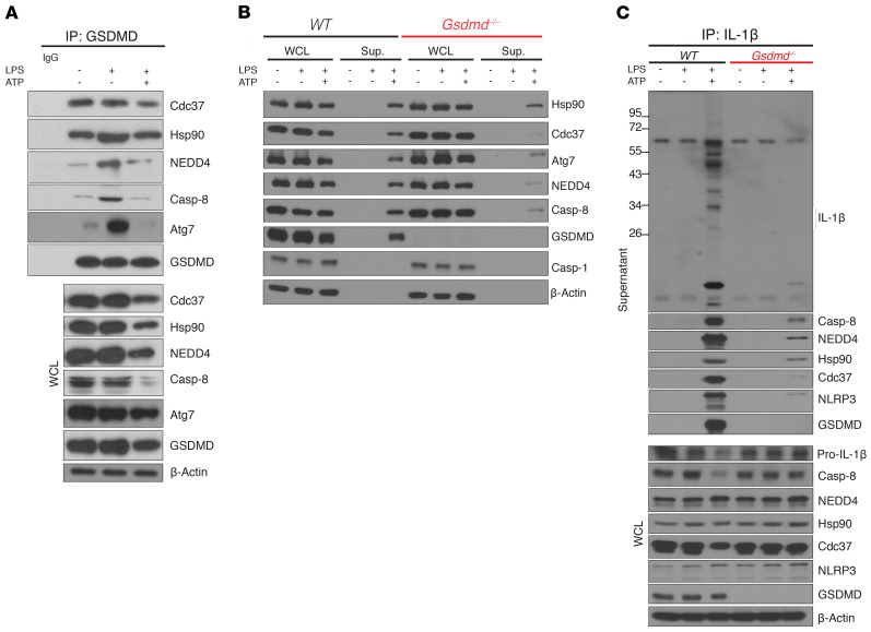
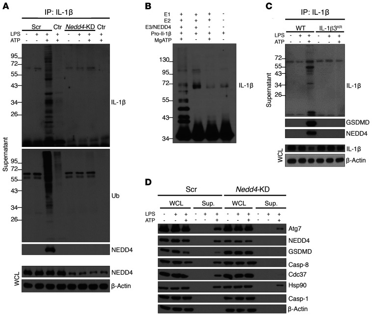
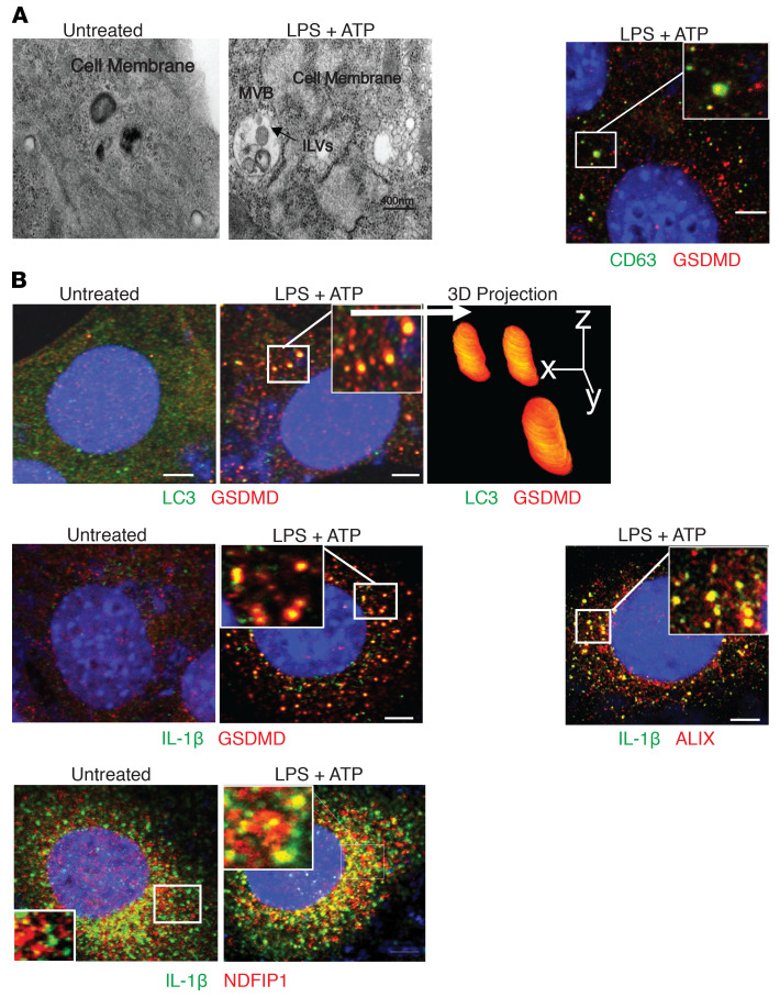
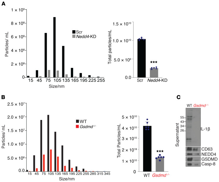
Gasdermin D (GSDMD) induces pyroptosis via the pore-forming activity of its N-terminal domain, cleaved by activated caspases associated with the release of IL-1β. Here, we report a nonpyroptotic role of full-length GSDMD in guiding the release of IL-1β-containing small extracellular vesicles (sEVs) from intestinal epithelial cells (IECs). In response to caspase-8 inflammasome activation, GSDMD, chaperoned by Cdc37/Hsp90, recruits the E3 ligase, NEDD4, to catalyze polyubiquitination of pro-IL-1β, serving as a signal for cargo loading into secretory vesicles. GSDMD and IL-1β colocalize with the exosome markers CD63 and ALIX intracellularly, and GSDMD and NEDD4 are required for release of CD63+ sEVs containing IL-1β, GSDMD, NEDD4, and caspase-8. Importantly, increased expression of epithelial-derived GSDMD is observed both in patients with inflammatory bowel disease (IBD) and those with experimental colitis. While GSDMD-dependent release of IL-1β-containing sEVs is detected in cultured colonic explants from colitic mice, GSDMD deficiency substantially attenuates disease severity, implicating GSDMD-mediated release of IL-1β sEVs in the pathogenesis of intestinal inflammation, such as that observed in IBD.

Keywords: Cytokines; Gastroenterology; Inflammation; Inflammatory bowel disease; Innate immunity.


Conflict of interest statement

Conflict of interest: The authors have declared that no conflict of interest exists.

Figures

Figure 1
Figure 1. GSDMD localizes to the colonic epithelium and its deficiency protects from DSS-induced colitis.
Colitis was induced in cohoused, sex-matched Gsdmd–/– (n = 10) and control Gdsmd+/– (n = 11) littermate mice by administration of 3% DSS in drinking water. Severity of colitis, assessed by (A) weight loss, (B) disease activity index (DAI), and (C) colon length (decreased length indicates increased inflammation). (D) Representative images of H&E-stained colons from DSS-treated mice (left), with histologic analysis of colitis performed on day 9 (right). Scale bars: 50 µM. (E) Representative images of immunofluorescence staining for CD4, F4/80, and LyG6 on frozen sections of inflamed colons taken at day 9 (left), with number of infiltrating immune cells enumerated and averaged over 10 HPF/section (right). Original magnification, ×40. (F) RT-PCR showing relative differences of indicated proinflammatory gene transcripts in colon tissues harvested on day 9 from experimental mice. Data are presented as fold induction over the mean of Gdsmd+/– group. (G) Representative IHC images showing immunolocalization of GSDMD to IECs in colons of DSS-challenged Gsdmd+/– control mice, with total absence in Gsdmd–/– littermates. Scale bars: 50 µM. Data are presented as mean ± SEM with *P < 0.05, ***P < 0.0001 by Student’s t test. All experiments were repeated twice and yielded consistent results.
Figure 2
Figure 2. Increased expression of GSDMD in biopsies and isolated IECs from gut mucosa of IBD patients compared with healthy controls.
(A) Analysis of 5 different IBD gene array expression data sets (Supplemental Table 1) derived from intestinal mucosal biopsies from CD, UC, and healthy controls (HC) displayed as box plots (25th, 50th, and 75th quantiles shown), with differences expressed as FDR between IBD patients and healthy controls. Act, active disease; Act-I, active involved area; Act-NI, active noninvolved area; Remiss., remission from IBD. (B) Representative Western blots of whole-cell lysates (WCLs) showing differences in GSDMD protein levels in freshly isolated IECs from IBD patients versus controls (n = 4),with relative densitometric values calculated as ratio of GSDMD/GAPDH and reported as mean ± SEM; **P < 0.005 versus control (Cont.) by 2-tailed unpaired Student’s t test.
Figure 3
Figure 3. Prevalence of GSDMD expression in specific epithelial subpopulations from inflamed mucosa of IBD patients.
(A) t-SNE plots after scRNa-Seq of colonic IECs depicting expression and distribution of GSDMD, with (B) bar plots showing GSDMD in epithelial subpopulations from UC patients (inflamed and noninflamed) and healthy controls (HC) (n = 3). DESeq2 R package was used to compute library size factors, normalize data, and perform differential expression analysis using negative binomial Wald’s test.
Figure 4
Figure 4. GSDMD-dependent release of polyubiquitinated IL-1β from colonic IECs.
Representative Western blots of (A) colon explant cultures from untreated (Utx) and DSS-treated (day 9) Gsdmd+/– and Gsdmd–/– mice, and of (BE) WT and GSDMD-deficient (Gsdmd–/–) YAMC cells; mature (m)IL-1β, full-length (FL) GSDMD, and ubiquitin (Ub). Supernatants were subjected to IP with anti–IL-1β and probed with anti–IL-1β (B) (WCLs were directly analyzed), or subjected to IP with anti–IL-1β under nondenaturing and denaturing conditions and probed with anti-Ub (C). (D) WCLs were directly analyzed. (E) Total protein was analyzed in supernatants after extraction (WCLs were directly analyzed). All experiments were repeated 3 times and yielded consistent results.
Figure 5
Figure 5. Polyubiquitinated IL-1β from colonic IECs is released via a caspase-8–dependent nonlytic pathway.
(A and B) Gsdmd–/– YAMC cells were stably restored with either WT or D276A GSDMD (GsdmdD276A), and stimulated as indicated. (A) Supernatants from stimulated cells were subjected to IP with anti–IL-1β (WCLs were directly analyzed). (B) Total protein in cell supernatants was extracted and analyzed, alongside WCLs. (C) Pore-forming ability was assessed by kinetic analysis of propidium iodide (PI) uptake in LPS- and ATP-stimulated WT and Gsdmd–/– YAMC cells, with triton treatment used as a positive control. Error bars are shown as SEM of technical replicates (n = 3). (D) IL-1β–deficient (IL-1β–KO) and WT control YAMC cells were left untreated or stimulated with LPS followed by ATP, and analyzed for indicated genes. (E) WT YAMC cells transduced with scramble (Scr) or caspase-8 targeting (Casp8 KD) shRNA were subjected to co-IP with anti–IL-1β. Precipitated supernatant proteins and WCLs were analyzed by Western blot. (F) WT YAMC cells were subjected to co-IP with anti-GSDMD and Western analysis as indicated. All experiments were repeated 3 times and yielded consistent results. All error bars show SEM with *P < 0.05, **P < 0.01, ***P < 0.001 by 2-tailed unpaired Student’s t test.
Figure 6
Figure 6. Nonlytic release of IL-1β from IEC requires assembly of a secretory complex guided by chaperoned pool of GSDMD.
Representative Western blots of (A) lysates from unstimulated, LPS-treated, and LPS+ATP-stimulated WT YAMC cells subjected to IP with anti-GSDMD and probed for indicated proteins, (B) WT and Gsdmd–/– YAMC cells either unstimulated or LPS/LPS+ATP-stimulated, with WCL and protein extracts from supernatants probed for indicated proteins, and (C) supernatants from WT and Gsdmd–/– YAMC cells with indicated treatments subjected to IP with anti–IL-1β, and probed for indicated proteins. All experiments were repeated at least 3 times and yielded consistent results.
Figure 7
Figure 7. Polyubiquitination of pro–IL-1β by NEDD4 facilitates its secretion via caspase-8–dependent GSDMD-guided nonlytic pathway.
YAMC cells were lentivirally transduced with either scramble (Scr) or NEDD4-targeting shRNA (Nedd4-KD), and stable clones treated as indicated; cells with no guide RNA were used as controls (Ctr). Representative Western blots of (A) total protein from supernatants pulled down with anti–IL-1β, and (B) in vitro ubiquitination assays with recombinant human proteins E1 (Ube1), E2 (UbcH5c/Ube2D3), E3 (NEDD4), and IL-1β as a substrate followed by mass spectometry analysis (Supplemental Table 3). (C) IL-1β–deficient YAMC cells were restored with either WT or mutant (IL-1β3KA) cDNA. Cells were treated as indicated, and total protein from supernatants was directly analyzed or subjected to IL-1β IP followed by Western blotting. (D) WCLs and protein extracts from supernatants were probed for indicated proteins. Except for mass spectrometry analysis, all experiments were repeated 3 times and yielded consistent results.
Figure 8
Figure 8. Characterization of GSDMD-associated proteins released from isolated IEC-derived sEVs.
WT and Gsdmd–/– YAMC cells were left untreated or stimulated with LPS+ATP. (A) Total concentrations of particles (15-285 nm), and (B) average diameters of particles, in indicated supernatants. Adjusted P values were calculated by Turkey’s multiple comparisons test (n = 3). (C) Representative images of transmission electron microscopic scanning of counterstained EVs from supernatants of untreated and LPS+ATP-treated cells, with (D) examples of EVs of varying sizes. Scale bars: 100 nM. (E) Representative Western blots of EVs from indicated supernatants after enrichment by size exclusion column-based fractionation. All experiments were repeated 3 times and yielded consistent results. Electron microscopy was performed on samples from 2 independent repeats, which generated vesicles of consistent morphology and range of diameters.
Figure 9
Figure 9. GSDMD-guided secretion of IL-1β requires vesicle biogenesis.
Representative photomicrographs of WT YAMC cells untreated or treated with LPS+ATP imaged by (A) transmission electron microscopy (left), and by confocal microscopy after staining for (B) CD63 (green) and GSDMD (red) (right), and (B) for indicated proteins, with 3D projections based on Z stack scanning of 100 planes of boxed region. All immunofluorescence imaging was repeated over 3 biological replicates in 3 independent experiments, which yielded consistent pattern of staining. Electron microscopy was performed on samples from 2 independent repeats. Confocal microscopy was performed 3 times and revealed consistent morphology. Scale bars: 10 µM (A), 15 µM (B).
Figure 10
Figure 10. GSDMD and NEDD4 mediate the release of a GSDMD/NEDD4/IL-1β complex via sEVs.
(A) YAMC cells transduced with either scramble shRNA (Scr) or shRNA targeting Nedd4 (Nedd4-KD) were stimulated with LPS, and supernatants were concentrated and subjected to size-exclusion columns to isolate exosomes, followed by nanoparticle tracking analysis with ZetaView for size distribution and total particle concentration (n = 5). (B) Supernatants from cultured colon explants from DSS-treated (day 9) WT and Gsdmd–/– mice were fractioned by size-exclusion columns to enrich for EVs followed by nanoparticle tracking analysis for size distribution and total particle concentration (n = 6), and evaluated for (C) indicated proteins, as shown by representative Western blots. ***P < 0.0001 by 2-tailed unpaired Student’s t test. All experiments were repeated 3 times and yielded consistent results.

References

    1. de Souza HS, Fiocchi C. Immunopathogenesis of IBD: current state of the art. Nat Rev Gastroenterol Hepatol. 2016;13(1):13–27. doi: 10.1038/nrgastro.2015.186. - DOI - PubMed
    1. Levy M, et al. Microbiota-modulated metabolites shape the intestinal microenvironment by regulating NLRP6 inflammasome signaling. Cell. 2015;163(6):1428–1443. doi: 10.1016/j.cell.2015.10.048. - DOI - PMC - PubMed
    1. Seo SU, et al. Distinct commensals induce interleukin-1β via NLRP3 inflammasome in inflammatory monocytes to promote intestinal inflammation in response to injury. Immunity. 2015;42(4):744–755. doi: 10.1016/j.immuni.2015.03.004. - DOI - PMC - PubMed
    1. Sorbara MT, Girardin SE. Mitochondrial ROS fuel the inflammasome. Cell Res. 2011;21(4):558–560. doi: 10.1038/cr.2011.20. - DOI - PMC - PubMed
    1. Zhao Y, et al. The NLRC4 inflammasome receptors for bacterial flagellin and type III secretion apparatus. Nature. 2011;477(7366):596–600. doi: 10.1038/nature10510. - DOI - PubMed

Publication types

MeSH terms