. 2020 Aug 20;48(14):7958-7972.
doi: 10.1093/nar/gkaa532.

Asymmetric dimerization of adenosine deaminase acting on RNA facilitates substrate recognition

Affiliations

Asymmetric dimerization of adenosine deaminase acting on RNA facilitates substrate recognition

Alexander S Thuy-Boun et al. Nucleic Acids Res. .

Abstract

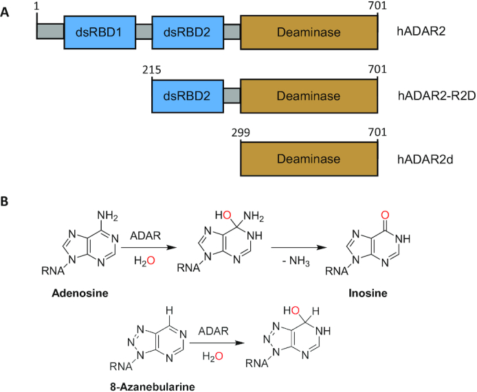
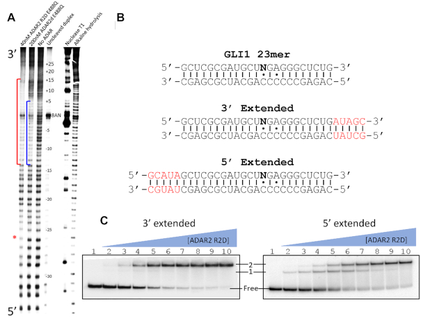
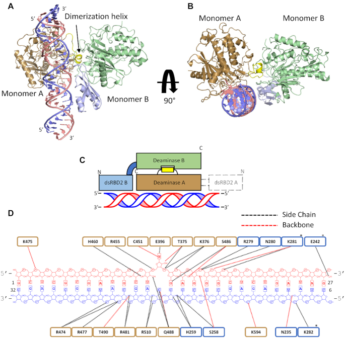
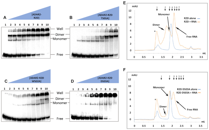
Adenosine deaminases acting on RNA (ADARs) are enzymes that convert adenosine to inosine in duplex RNA, a modification that exhibits a multitude of effects on RNA structure and function. Recent studies have identified ADAR1 as a potential cancer therapeutic target. ADARs are also important in the development of directed RNA editing therapeutics. A comprehensive understanding of the molecular mechanism of the ADAR reaction will advance efforts to develop ADAR inhibitors and new tools for directed RNA editing. Here we report the X-ray crystal structure of a fragment of human ADAR2 comprising its deaminase domain and double stranded RNA binding domain 2 (dsRBD2) bound to an RNA duplex as an asymmetric homodimer. We identified a highly conserved ADAR dimerization interface and validated the importance of these sequence elements on dimer formation via gel mobility shift assays and size exclusion chromatography. We also show that mutation in the dimerization interface inhibits editing in an RNA substrate-dependent manner for both ADAR1 and ADAR2.


Figures

Figure 1.
Figure 1.
(A) Domain map of hADAR2 and deletion fragments. (B) Reaction mechanism of ADAR2 showing intermediate and 8-azanebularine (8-AN) hydrate.
Figure 2.
Figure 2.
MPE•Fe footprinting and electrophoretic mobility shift assay (EMSA) of 8-AN containing RNA. (A) Polyacrylamide gel of 5′ 32P labeled RNA fragments resulting from limited MPE•Fe cleavage. Red brackets represent regions in lane 1 that show strong cleavage protection by hADAR2-R2D, the blue brackets represent regions in lane 2 that show strong cleavage protection by hADAR2d. Red asterisk indicates site of protection by ADAR2-R2D 5′ to editing site. (B) Sequence of GLI1 RNA used for gel shift assay. 8-AN is represented as N. Extensions from 23 bp duplex ligand of hADAR2d are shown in red. (C) Gel shift of hADAR2-R2D with 3′ extended or 5′ extended RNA duplex. Protein concentrations are as follows: lane 1: no protein, lanes 2–10: 0.25, 0.5, 1, 2, 4, 8, 16, 32, 64 nM.
Figure 3.
Figure 3.
Structure of hADAR2-R2D E488Q bound to the GLI1 32 bp RNA at 2.8 Å resolution. (A) View of the structure perpendicular to the dsRNA helical axis. (B) View of the structure along the dsRNA helical axis. (C) Cartoon schematic of the asymmetric protein dimer. Colors for ADAR2 domains and RNA strands correspond to those used in A and B. Electron density for dsRBD2 on monomer A was not resolved. (D) Summary of contacts between hADAR2-R2D E488Q and GLI1 32 bp RNA duplex. Brown represent RNA contacts to deaminase A; blue represents RNA contacts to dsRBD2 of monomer B. Asterisk represent potential contacts that lack full side-chain electron density.
Figure 4.
Figure 4.
ADAR contacts observed in protein dimer. (A) Protein–protein contacts within dimerization interface. Monomer A is primarily colored brown, the dimerization helix of Monomer A is colored yellow, Monomer B is colored green. (B) Contacts between dsRBD2 B and RNA duplex. dsRBD B is labeled in light blue, 8-AN containing RNA strand is labeled in light pink, the complementary strand is labeled in blue. (C) Representative sequence alignment for ADARs showing sequence conservation in dimerization helix. Conserved residues contained within ADAR2′s dimerization helix are highlighted red.
Figure 5.
Figure 5.
Representative gel shift and gel filtration of hADAR2-R2D E488Q mutants with and without 3′ extended 8-AN RNA. Gel shift and gel filtration experiments were all performed with the E488Q mutation in addition to any other point mutations listed. (A–D) Protein concentrations are as follows: 1: no protein, 2: 0.25 nM, 3: 0.5 nM, 4: 1 nM, 5: 2 nM, 6: 4 nM, 7: 8 nM, 8: 16 nM, 9: 32 nM, 10: 64 nM. The band of RNA retained within the well arises from protein–RNA aggregation. (E, F) UV trace of hADAR2-R2D eluted from a Superdex Increase 5/150 GL. The elution volumes of each peak for the R2D ± 3′ extend 8-AN RNA runs are as follows: (*) 1.17 ml, dimer 1.71 ml, monomer 1.86 ml, R2D alone 1.91 ml. The elution volumes of each peak for the R2D D503A ± 3′ extended 8-AN RNA runs are as follows: (*) 1.19 ml, monomer 1.88 ml, R2D D503A alone 1.91 ml. The elution peaks of molecular weight standards are indicated and numbered 1–7 at top. The sizes and retention volumes are as follows: (1) thyroglobulin (bovine) 670 kDa, 1.19 ml, (2) γ-globulin (bovine) 158 kDa, 1.59 ml, (3) bovine serum albumin 66 kDa, 1.81 ml, (4) ovalbumin (chicken) 44 kDa, 1.95 ml, (5) carbonic anhydrase 29 kDa, 2.12 ml, (6) myoglobin (horse) 17 kDa, 2.21 ml, (7) cytochrome c 12.4 kDa, 2.28 ml. The observed molecular weight corresponding to each protein species can be found in Supplementary Table S3. The peaks designated by an asterisk correspond to protein and RNA eluted within the void volume, representing aggregation.
Figure 6.
Figure 6.
Deamination assays with 100 nM ADAR and 10 nM 5-HT2cR D site substrate. (A) Local predicted secondary structure of 5-HT2cR pre-mRNA used for in vitro deamination. (B) Deamination kinetics of ADAR2-RD E488Q and dimerization mutants on 5-HT2cR substrate (D-site). Conditions for in vitro deaminations are stated in Table 2. Error bars represent standard deviation (n = 3 technical replicates).
Figure 7.
Figure 7.
Editing levels of endogenous substrates in HEK293T cells with or without overexpression of ADARs. (A) Editing by full length human ADAR2 wild type (A2WT) and dimerization mutants and (B) by human ADAR1 p110 wild type (A1 p110) and A1 p110 D1023A mutant. Cells were transfected with 750 ng pcDNA 3.1 containing A2WT or A1 p110 gene and lysed after 48 h. Endogenous editing was measured from cells transfected with empty vector. Asterisks indicate no detected editing. Error bars represent standard deviation (n = 3 biological replicates).

Similar articles

Cited by

References

    1. Grosjean H., Grosjean H.. 2005; Springer.
    1. Bass B.L. RNA editing by adenosine deaminases that act on RNA. Annu. Rev. Biochem. 2002; 71:817–846. - PMC - PubMed
    1. Nishikura K. Functions and regulation of RNA editing by ADAR deaminases. Annu. Rev. Biochem. 2010; 79:321–349. - PMC - PubMed
    1. Rueter S.M., Dawson T.R., Emeson R.B.. Regulation of alternative splicing by RNA editing. Nature. 1999; 399:75. - PubMed
    1. Yeo J., Goodman R.A., Schirle N.T., David S.S., Beal P.A.. RNA editing changes the lesion specificity for the DNA repair enzyme NEIL1. Proc. Natl. Acad. Sci. U.S.A. 2010; 107:20715–20719. - PMC - PubMed

Publication types