. 2020 Jun 24;12(6):585.
doi: 10.3390/pharmaceutics12060585.

Development of ErbB2-Targeting Liposomes for Enhancing Drug Delivery to ErbB2-Positive Breast Cancer

Affiliations

Development of ErbB2-Targeting Liposomes for Enhancing Drug Delivery to ErbB2-Positive Breast Cancer

Sho Ueno et al. Pharmaceutics. .

Abstract

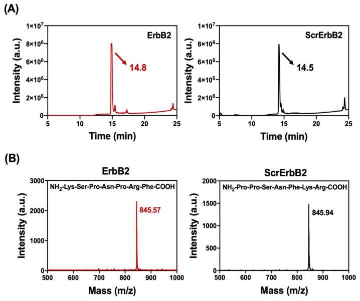
ErbB2 is a type of receptor tyrosine kinase, which is known to be involved in tumorigenesis, tumor aggressiveness, and clinical outcome. ErbB2-targeting therapy using therapeutic antibodies has been successful in breast cancer treatment. However, the need for repeated treatments and the high cost are major disadvantages with monoclonal antibody therapies. Compared with antibodies, peptides are cheap, relatively stable, and have low immunogenicity. We have developed a highly specific cancer-targeting drug delivery system using a targeting peptide to maximize the therapeutic efficiency of rapamycin and to help prevent drug resistance in ErbB2-positive breast cancer. Physicochemical characterization confirmed the successful construction of ErbB2-targeting liposomes (ErbB2Lipo). A comparison of a scrambled peptide (ScrErbB2) with the ErbB2-targeting peptide confirmed that these peptides had similar properties except for the targeting ability. The ErbB2Lipo exhibited higher delivery efficiency in ErbB2 positive BT-474 cells than non-targeting liposomes conjugated with ScrErbB2 (ScrErbB2Lipo). This peptide-targeting strategy has the potential to improve the efficacy of chemotherapy in ErbB2-positive cancers.

Keywords: breast cancer therapy; drug delivery system; immunoliposome; mTOR inhibitor; targeted therapy; targeting peptide.


Conflict of interest statement

The authors declare no conflict of interest.

Figures

Figure 1
Figure 1
(A) The absorbance measured at 220 nm is representative of peptide bonds. A distinct peak confirmed the purity of the peptide synthesized by solid-phase peptide synthesis. The ErbB2 and ScrErbB2 are indicated by arrows. (B) Matrix-assisted laser desorption ionization-time-of-flight (MALDI-TOF) mass spectroscopy spectra of purified peptides. Samples were collected at 14–15 min by high-performance liquid chromatography (HPLC). The peaks corresponding to [M + H]+ of the peptides ErbB2 (KSPNPRF) and ScrErbB2 (PPSNFKR) had m/z values of 845.57 and 845.94, respectively (m/z expected: 844.45).
Figure 2
Figure 2
The 1H NMR spectra of (a) DSPE-PEG-ErbB2, (b) DSPE-PEG-NH2, and (c) the ErbB2 peptide. The red dashed squares indicate the chemical shifts of the methine and methylene protons of the ErbB2 peptide and the methylene protons of DSPE-PEG-NH2.
Figure 3
Figure 3
Physicochemical characteristics of the liposomes. (A) The particle size, polydispersity index (PDI), and zeta-potential values were measured using dynamic light scattering. (B) Transmission electron microscope images of BareLipo, ErbB2Lipo, and ScrErbB2Lipo.
Figure 4
Figure 4
Analysis of binding between rhodamine B-labeled liposomes and breast cancer cells using flow cytometry. BT-474 and MDA-MB-231 cells were treated with control, ScrErbB2Lipo, and ErbB2Lipo. (A) Plots of flow cytometry results and (B) quantitative analysis of Figure 4A.
Figure 5
Figure 5
Cellular internalization of the ErbB2Lipo. Representative confocal microscopy images of (A) BT-474 and (B) MDA-MB-231 cells after incubation for 3 h. Cell nuclei was stained with Hoechst 33342 (blue). Rhodamine B (red) indicated the presence of liposomes. Scale bar: 10 μm.
Figure 6
Figure 6
The cytotoxicity of ScrErbB2Lipo and ErbB2Lipo after 72 h in (A) BT-474 and (B) MDA-MB-231 cells. Results are presented as mean ± SEM (n = 8).

References

    1. DeSantis C.E., Ma J.M., Gaudet M.M., Newman L.A., Miller K.D., Sauer A.G., Jemal A., Siegel R.L. Breast cancer statistics, 2019. Ca-Cancer J. Clin. 2019;69:438–451. doi: 10.3322/caac.21583. - DOI - PubMed
    1. Harari D., Yarden Y. Molecular mechanisms underlying ErbB2/HER2 action in breast cancer. Oncogene. 2000;19:6102–6114. doi: 10.1038/sj.onc.1203973. - DOI - PubMed
    1. Tai W., Mahato R., Cheng K. The role of HER2 in cancer therapy and targeted drug delivery. J. Control. Release. 2010;146:264–275. doi: 10.1016/j.jconrel.2010.04.009. - DOI - PMC - PubMed
    1. Tovey S.M., Brown S., Doughty J.C., Mallon E.A., Cooke T.G., Edwards J. Poor survival outcomes in HER2-positive breast cancer patients with low-grade, node-negative tumours. Br. J. Cancer. 2009;100:680–683. doi: 10.1038/sj.bjc.6604940. - DOI - PMC - PubMed
    1. Masoud V., Pages G. Targeted therapies in breast cancer: New challenges to fight against resistance. World J. Clin. Oncol. 2017;8:120–134. doi: 10.5306/wjco.v8.i2.120. - DOI - PMC - PubMed