. 2020 Jun 24;9(6):549.
doi: 10.3390/antiox9060549.

Reversible Oxidative Modifications in Myoglobin and Functional Implications

Affiliations

Reversible Oxidative Modifications in Myoglobin and Functional Implications

Mark H Mannino et al. Antioxidants (Basel). .

Abstract

Myoglobin (Mb), an oxygen-binding heme protein highly expressed in heart and skeletal muscle, has been shown to undergo oxidative modifications on both an inter- and intramolecular level when exposed to hydrogen peroxide (H2O2) in vitro. Here, we show that exposure to H2O2 increases the peroxidase activity of Mb. Reaction of Mb with H2O2 causes covalent binding of heme to the Mb protein (Mb-X), corresponding to an increase in peroxidase activity when ascorbic acid is the reducing co-substrate. Treatment of H2O2-reacted Mb with ascorbic acid reverses the Mb-X crosslink. Reaction with H2O2 causes Mb to form dimers, trimers, and larger molecular weight Mb aggregates, and treatment with ascorbic acid regenerates Mb monomers. Reaction of Mb with H2O2 causes formation of dityrosine crosslinks, though the labile nature of the crosslinks broken by treatment with ascorbic acid suggests that the reversible aggregation of Mb is mediated by crosslinks other than dityrosine. Disappearance of a peptide containing a tryptophan residue when Mb is treated with H2O2 and the peptide's reappearance after subsequent treatment with ascorbic acid suggest that tryptophan side chains might participate in the labile crosslinking. Taken together, these data suggest that while exposure to H2O2 causes Mb-X formation, increases Mb peroxidase activity, and causes Mb aggregation, these oxidative modifications are reversible by treatment with ascorbic acid. A caveat is that future studies should demonstrate that these and other in vitro findings regarding properties of Mb have relevance in the intracellular milieu, especially in regard to actual concentrations of metMb, H2O2, and ascorbate that would be found in vivo.

Keywords: ditryptophan; dityrosine; myoglobin; peroxidase; protein aggregation.


Conflict of interest statement

The authors declare no conflict of interest.

Figures

Figure 1
Figure 1
Pre-treatment with H2O2 increases MetMb peroxidase activity in a substrate-dependent manner. MetMb (111 μM, pH 5.9) was untreated (UT) or was pre-reacted with 50 μM H2O2 for 15 min at (A) pH 7.4 (n = 12/group, * p < 0.05) and (B) pH 5.9 (n = 7/group, * p < 0.05) before 2 μL of this solution was added to a 200 μL reaction mixture on a 96-well plate containing 250 μM ascorbic acid and 200 μM H2O2. (C) metMb was reacted with H2O2 at pH 5.9 as described for (B), and peroxidase activity was measured using 500 μM TMB and 200 μM H2O2 (n = 10/group). (D) MetMb (222 μM) was pre-reacted with 100 μM H2O2 for 30 min before 1 μL of this solution was added to a 200 μL reaction mixture containing 250 μM ascorbic acid and 200 μM H2O2 at pH 8.5. * p ≤ 0.05, n = 12/group.
Figure 2
Figure 2
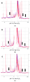
Analysis of heme-protein crosslinks by 700 MHz 1H NMR spectra. (AB) MetMb (555 µM, pH 5.9) was reacted with 110 µM H2O2 (A) or 1.1 mM H2O2 (B) for 10 min before adding 3 mM NaCN to generate low-spin Mb. Sample preparation and 1H NMR procedures are described in the methods section. (C) Same as (A) and (B), only metMb was reacted with 110 µM H2O2 at pH 7.4. Data for untreated metMb are in red, and data for metMb pretreated with H2O2 are in blue. Arrows indicate novel peaks in the heme region for H2O2-treated metMb.
Figure 3
Figure 3
Reaction of metMb with H2O2 increases peroxidase activity as visualized on polyacrylamide gels. metMb (555 μM, pH 5.9) was incubated in the absence or presence of 800 μM H2O2 for 15 min before performing SDS-PAGE and (A) heme-peroxidase activity stains (right) as described in methods. (B) Quantitation of the peroxidase product at the 17 kDa band, n = 3/group, * p < 0.01.
Figure 4
Figure 4
Mb-X species possess unique peroxidase activities compared to metMb. (A) Mb-X peroxidase activity was measured using ascorbic acid (Asc, 250 μM), resveratrol (Resv, 125 μM), NADH (250 μM) and TMB (500 μM) and 200 μM H2O2 at pH 6.1. The activity plots for all substrates except ascorbic acid overlap, so they are not all visible. n = 6/group, * p <0.05 vs. all other groups. (B) Mb-X concentration was estimated as described in the methods section. Peroxidase activity with ascorbic acid was then measured with 250 μM ascorbic acid and 200 μM H2O2 at pH 6.1. * p ≤ 0.05 compared to metMb, n = 3/group. (C) Mb-X peroxidase activity using caffeic acid (125 μM), TMB (500 μM) and NAD(P)H (250 μM) and 200 μM H2O2, pH 6.1 both in the presence and absence of 50 μM ascorbic acid. * p ≤ 0.05 compared to control without ascorbic acid, n = 3/group. (D) Mb-X peroxidase activity was measured at pH 7.4 and 6.1 using 250 μM ascorbic acid and 200 μM H2O2 (n = 3/group).
Figure 5
Figure 5
Heme-dependent reaction of H2O2-oxidized metMb with ascorbic acid is sufficient to reverse heme-protein crosslinks. (A) MetMb (111 μM, pH 6.1) was incubated in the absence or presence of 300 μM H2O2 for 10 min prior to adding 833 μM ascorbic acid for an additional 10 min and performing SDS-PAGE followed by (A) a heme peroxidase stain. (B) Quantitation of the peroxidase product at the 17 kDa band, n = 5/group, * p < 0.001. (C) Acid-butanone heme extraction was performed on metMb that was either untreated, reacted with H2O2 alone for 10 min (H2O2) or treated with ascorbic acid after 10 min of H2O2 oxidation. Free heme was assessed by absorbance at 398 nm. * p ≤ 0.05 compared to untreated control; ¥ p ≤ 0.05 compared to H2O2-only; n = 3/group.
Figure 6
Figure 6
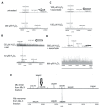
Matrix assisted laser desorption mass spectrometry-time of flight (MALDI-TOF) mass spectrometric analysis of ascorbic acid-mediated reversal of heme-protein crosslinks in H2O2-reacted metMb. (A) (Bottom spectra) MetMb tryptic digests that were reacted with 100 μM H2O2 displayed loss of the N-terminal peptide (1815.9 m/z) indicated by arrows in the untreated MetMb (top spectra). (BD) Tryptic digests of H2O2-reacted metMb that was then treated with ascorbic acid revealed the re-appearance of the N-terminal peptide. (E). MetMb monomer (bottom) and dimer (top) tryptic digests that were reacted with 300 µM H2O2 and analyzed with MALDI-TOF MS. The arrow indicates a peak at 1993 m/z, which suggests presence of a heme crosslinked to a peptide containing the distal histidine of Mb helix E.
Figure 7
Figure 7
Substrate competition for metMb peroxidase activity. MetMb peroxidase activity with NADH (A) or NADPH (B) was measured in the presence of varying concentrations of ascorbic acid. * p ≤ 0.05 compared to NAD(P)H without ascorbic acid present. n = 6/group in (A) and n = 11–12/group in (B).
Figure 8
Figure 8
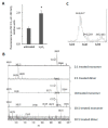
Reaction of horse metMb with H2O2 results in dityrosine formation. (A) Fluorescence of metMb was measured before and after oxidation with H2O2 using excitation and emission wavelengths of 290 nm and 400 nm, respectively. * p ≤ 0.005 compared to untreated, n = 6/group. (B) MALDI-TOF analysis of metMb tryptic digests revealed that the tryptic peptide containing tyrosine 103 (1885 m/z) was absent in the H2O2-treated dimer. From bottom: dimer from 222 μM Mb reacted with 2.2 mM H2O2; monomer from 222 μM Mb reacted with 2.2 mM H2O2; untreated monomer; 222 μM Mb dimer reacted with 444 μM H2O2; 222 μM Mb monomer reacted with 444 μM H2O2. Arrows indicate peaks for 1885 m/z. (C) MALDI-TOF analysis of oxidized metMb dimers indicate the presence of a Y103-Y146 dityrosine cross-linked peptide with a m/z of 3436.3.
Figure 9
Figure 9
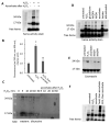
Treatment with ascorbic acid reverses crosslinking of Mb dimers. (A) Heme peroxidase activity stain of metMb that was untreated, H2O2-treated, or H2O2-treated followed by exposure to ascorbic acid. (B) Fluorescence of metMb treated as for panel A was assessed at excitation 290 nm and emission 400 nm, which correspond to the fluorescence characteristics of dityrosine [17], n = 8/group, * p < 0.001 vs untreated and group treated with peroxide and then ascorbic acid, † p = 0.05 vs untreated. (C) Western blot of ascorbic acid-treated oxidized metMb using an anti-dityrosine antibody. (D) Heme stain of H2O2-reacted metMb that was then treated with various reducing substrates. (E) Coomassie stain of Mb exposed to H2O2 and then reducing agents. (F) Heme stain of oxidized metMb that was denatured by heating for 10 min at 90 °C or by adding SDS-PAGE sample loading buffer prior to the addition of ascorbic acid.
Figure 10
Figure 10
Role of tyrosine residues in regulating oxidatively-modified metMb species. (A) Peroxidase activity assay of metMb that had been treated with H2O2 alone, N-acetylimidazole (NAI) alone or H2O2 and then NAI (as described in methods and materials) using 200 μM H2O2 and 250 μM ascorbic acid at pH 6.1. * p ≤ 0.05 compared to untreated control, n = 6/group. Activity plots for H2O2 and NAI groups overlap, so they are not both visible. (B) Coomassie stains of the samples prepared in (A). Lanes 1–4: untreated, control metMb; H2O2-treated, control metMb; H2O2-treated control metMb + ascorbic acid; H2O2-treated control metMb + NADH. Lanes 5–8: same as (1–4) only using met Mb that had been reacted with H2O2 (pre-oxidized). Lanes 9–12: same as (1–4) and (5–8) only using NAI-treated metMb. Lanes 13–16: same only using H2O2-treated then NAI-treated metMb. Lanes showing that tyrosine-acetylated Mb cannot reverse Mb dimerization in the presence of ascorbic acid are indicated by arrows.

Similar articles

Cited by

References

    1. Stadtman E.R., Levine R.L. Free radical-mediated oxidation of free amino acids and amino acid residues in proteins. Amino Acids. 2003;25:207–218. doi: 10.1007/s00726-003-0011-2. - DOI - PubMed
    1. Boronat S., Garcia-Santamarina S., Hidalgo E. Gel-free proteomic methodologies to study reversible cysteine oxidation and irreversible protein carbonyl formation. Free Radic. Res. 2015;49:494–510. doi: 10.3109/10715762.2015.1009053. - DOI - PubMed
    1. Reeg S., Grune T. Protein oxidation in aging: Does it play a role in aging progression? Antioxid. Redox Signal. 2015;23:239–255. doi: 10.1089/ars.2014.6062. - DOI - PMC - PubMed
    1. Kim N.H., Jeong M.S., Choi S.Y., Kang J.H. Oxidative modification of cytochrome c by hydrogen peroxide. Mol. Cells. 2006;22:220–227. - PubMed
    1. Xiang W., Weisbach V., Sticht H., Seebahn A., Bussmann J., Zimmermann R., Becker C.M. Oxidative stress-induced posttranslational modifications of human hemoglobin in erythrocytes. Arch. Biochem. Biophys. 2013;529:34–44. doi: 10.1016/j.abb.2012.11.002. - DOI - PubMed

LinkOut - more resources