. 2020 Jun 29;11(1):259.
doi: 10.1186/s13287-020-01756-x.

Melatonin-stimulated MSC-derived exosomes improve diabetic wound healing through regulating macrophage M1 and M2 polarization by targeting the PTEN/AKT pathway

Affiliations

Melatonin-stimulated MSC-derived exosomes improve diabetic wound healing through regulating macrophage M1 and M2 polarization by targeting the PTEN/AKT pathway

Wei Liu et al. Stem Cell Res Ther. .

Abstract

Background: After surgery, wound recovery in diabetic patients may be disrupted due to delayed inflammation, which can lead to undesired consequences, and there is currently a lack of effective measures to address this issue. Mesenchymal stem cell (MSC)-derived exosomes (Exo) have been proven to be appropriate candidates for diabetic wound healing through the anti-inflammatory effects. In this study, we investigated whether melatonin (MT)-pretreated MSCs-derived exosomes (MT-Exo) could exert superior effects on diabetic wound healing, and we attempted to elucidate the underlying mechanism.

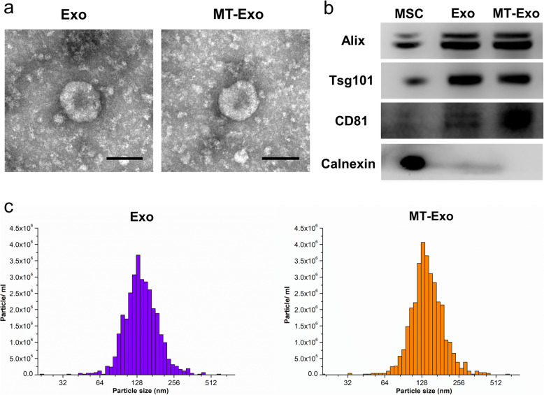
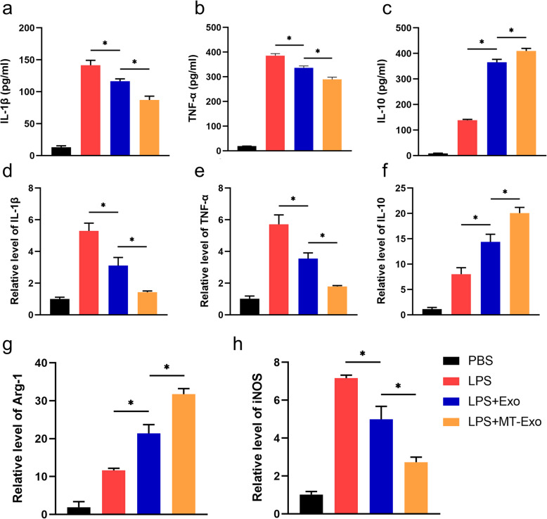
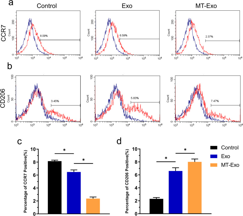
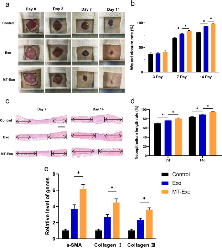
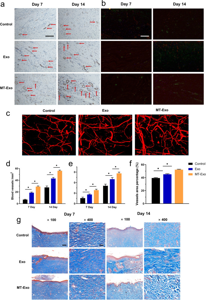
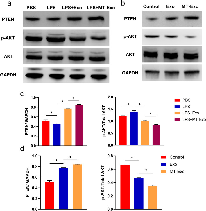
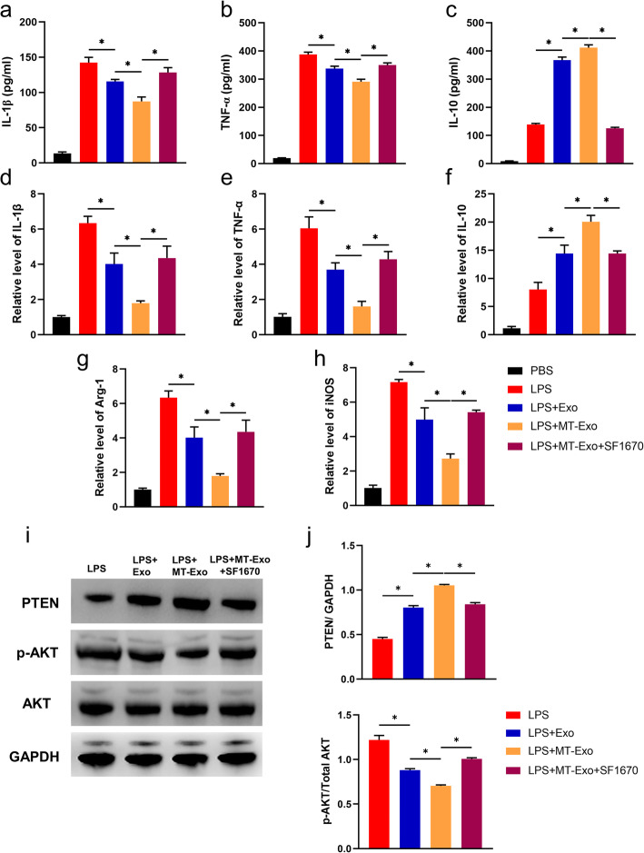
Methods: For the evaluation of the anti-inflammatory effect of MT-Exo, in vitro and in vivo studies were performed. For in vitro research, we detected the secreted levels of inflammation-related factors, such as IL-1β, TNF-α and IL-10 via ELISA and the relative gene expression of the IL-1β, TNF-α, IL-10, Arg-1 and iNOS via qRT-PCR and investigated the expression of PTEN, AKT and p-AKT by Western blotting. For in vivo study, we established air pouch model and streptozotocin (STZ)-treated diabetic wound model, and evaluated the effect of MT-Exo by flow cytometry, optical imaging, H&E staining, Masson trichrome staining, immunohistochemical staining, immunofluorescence, and qRT-PCR (α-SMA, collagen I and III).

Results: MT-Exo significantly suppressed the pro-inflammatory factors IL-1β and TNF-α and reduced the relative gene expression of IL-1β, TNF-α and iNOS, while promoting the anti-inflammatory factor IL-10 along with increasing the relative expression of IL-10 and Arg-1, compared with that of the PBS, LPS and the Exo groups in vitro. This effect was mediated by the increased ratio of M2 polarization to M1 polarization through upregulating the expression of PTEN and inhibiting the phosphorylation of AKT. Similarly, MT-Exo significantly promoted the healing of diabetic wounds by inhibiting inflammation, thereby further facilitating angiogenesis and collagen synthesis in vivo.

Conclusions: MT-Exo could promote diabetic wound healing by suppressing the inflammatory response, which was achieved by increasing the ratio of M2 polarization to M1 polarization through activating the PTEN/AKT signalling pathway, and the pretreatment of MT was proved to be a promising method for treating diabetic wound healing.

Keywords: Diabetic wound; Exosome; Inflammation; Macrophage polarization; Melatonin; Mesenchymal stem cell.


Conflict of interest statement

The authors declare no competing interests.

Figures

Fig. 1
Fig. 1
The characterization of Exo and MT-Exo. a The morphology of Exo and MT-Exo was analysed by TEM. Scale bar = 100 nm. b The surface markers (Alix, Tsg101, CD81 and Calnexin) of Exo and MT-Exo were evaluated by Western blotting. hBMSCs were used as a control and Calnexin was used as a negative control. c The diameter and concentration of exosomes were measured by NTA analysis (n = 3)
Fig. 2
Fig. 2
MT-Exo inhibited the inflammatory response by increasing the ratio of M2 polarization macrophages to M1 polarization in vitro. RAW264.7 cells were treated with PBS, LPS (100 ng/mL), LPS + Exo and LPS + MT-Exo for 24 h. ELISA was performed to detect the concentrations of a IL-1β, b TNF-α and c IL-10 from the supernatants. The relative gene expressions of d IL-1β, e TNF-α, f IL-10, g Arg-1 and h iNOS were detected (n = 3, *p < 0.05)
Fig. 3
Fig. 3
MT-Exo increased the ratio of M2 to M1 polarization in vivo. a, b Representative images of macrophage polarization of surface markers (CCR7 and CD206) of RAW264.7 by flow cytometry analysis (n = 3, *p < 0.05). c The quantitative analysis of percentage of CCR7 positive cells of the Control, Exo and MT-Exo group (n = 3, *p < 0.05). d The quantitative analysis of percentage of CD206 positive cells of the Control, Exo and MT-Exo group (n = 3, *p < 0.05)
Fig. 4
Fig. 4
MT-Exo expedited diabetic wound healing in vivo. a, b Optical images and related quantification of the wound closure rate of full-thickness dermal defects in the Control group, Exo group and MT-Exo group at day 0, 7 and 14 after the skin operation (n = 3, *p < 0.05, Scale bar = 10 mm). c, d H&E staining images and related quantification of total neoepithelium length in the Control group, Exo group and MT-Exo group at days 7 and 14 (n = 3, *p < 0.05). e The relative gene expression of the angiogenesis-related gene α-SMA and collagen synthesis-related genes Collagen I and III in the Control, Exo, and MT-Exo groups (n = 3, *p < 0.05)
Fig. 5
Fig. 5
MT-Exo improves angiogenesis and collagen synthesis in diabetic rats in vivo. a The assessment of the neovessels in the Control group, Exo group and MT-Exo group by α-SMA IHC at day 7 and 14 (red arrows display neovessels, Scale bar = 100 μm). b IF evaluation for CD31/α-SMA in the Control group, Exo group and MT-Exo group at days 7 and 14 (Scale bar = 100 μm). c The evaluation of neovasculars via the Microfil imaging method. d Quantification of the number of neovessels per field at day 7 and 14 by IHC (n = 3, *p < 0.05). e Quantification of the number of neovessels per field at day 7 and 14 by IF. f Quantification of the number of neovasculars per field at day 7 and 14 by Microfil. g Masson’s trichrome staining at day 7 and 14 post-operationally (Scale bar = 200 μm for 100× and 50 μm for 400×)
Fig. 6
Fig. 6
MT-Exo suppressed inflammation by activating the PTEN/AKT signalling pathway. a The expressions of PTEN, phosphorylation of AKT, AKT (total AKT) and GAPDH were tested in RAW264.7 cells after treatment with PBS, LPS (100 ng/mL), LPS + Exo and LPS + MT-Exo for 24 h by Western blotting. GAPDH was utilized as an internal reference. b The expressions of PTEN, phosphorylation of AKT, AKT (total AKT) and GAPDH were tested in vivo after treatment with PBS, Exo and MT-Exo by Western blotting. GAPDH was utilized as an internal reference. c The quantification of the greyscale values of PTEN/ GAPDH and p-AKT/AKT in vitro (n = 3, *p < 0.05). d The quantification of PTEN/ GAPDH and p-AKT/ AKT by Western blotting in vivo (n = 3, *p < 0.05)
Fig. 7
Fig. 7
PTEN inhibitor SF1670 antagonized the anti-inflammatory effect of MT-Exo. RAW264.7 cells were treated with PBS, LPS (100 ng/mL), LPS + Exo, LPS + MT-Exo and LPS + MT-Exo + SF1670 for 24 h. Then, ELISA was performed to detect the concentrations of a IL-1β, b TNF-α and c IL-10 from the supernatants. The relative gene expressions of d IL-1β, e TNF-α, f IL-10, g Arg-1 and h iNOS were detected via qRT-PCR (n = 3, *p < 0.05). i The expression of PTEN, phosphorylation of AKT, AKT (total AKT) and GAPDH were also tested. GAPDH was utilized as an internal reference. j The quantification of PTEN/GAPDH and p-AKT/AKT by Western blotting (n = 3, *p < 0.05)

References

    1. Mehta SK, Breitbart EA, Berberian WS, Liporace FA, Lin SS. Bone and wound healing in the diabetic patient. Foot Ankle Clin. 2010;15(3):411–437. - PubMed
    1. Chahal J, Stephen DJ, Bulmer B, Daniels T, Kreder HJ. Factors associated with outcome after subtalar arthrodesis. J Orthop Trauma. 2006;20(8):555–561. - PubMed
    1. Yan J, Tie G, Wang S, et al. Diabetes impairs wound healing by Dnmt1-dependent dysregulation of hematopoietic stem cells differentiation towards macrophages. Nat Commun. 2018;9(1):33. - PMC - PubMed
    1. Feng J, Dong C, Long Y, et al. Elevated Kallikrein-binding protein in diabetes impairs wound healing through inducing macrophage M1 polarization. Cell Commun Signal. 2019;17(1):60. - PMC - PubMed
    1. He R, Yin H, Yuan B, et al. IL-33 improves wound healing through enhanced M2 macrophage polarization in diabetic mice. Mol Immunol. 2017;90:42–49. - PubMed

Publication types