

Characterization of the role for cadherin 6 in the regulation of human endometrial receptivity
- PMID: 32600462
- PMCID: PMC7322878
- DOI: 10.1186/s12958-020-00624-w
Characterization of the role for cadherin 6 in the regulation of human endometrial receptivity
Abstract
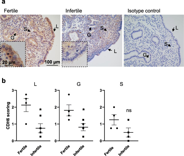
Background: The endometrial luminal epithelium is the first point of attachment of embryos during implantation. Failure of embryos to firmly adhere results in implantation failure and infertility. A receptive endometrial luminal epithelium is achieved through the expression of adhesion molecules in the mid-secretory phase and is a requirement for implantation. Cadherin 6 (CDH6) is an adhesion molecule localizing to the endometrial luminal epithelial cell surface in the mid-secretory/receptive phase and knockdown of CDH6 in the Ishikawa cells (receptive endometrial epithelial cell line) compromises cell integrity. However, there are no studies investigating the role of CDH6 on receptivity and infertility. This study aimed to investigate whether CDH6 is dysregulated in the endometrium of women with infertility during the receptive window and the effect of CDH6 on endometrial adhesion and receptivity.
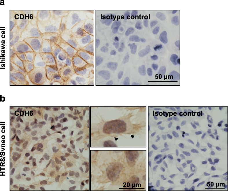
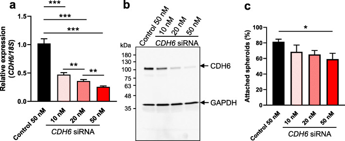
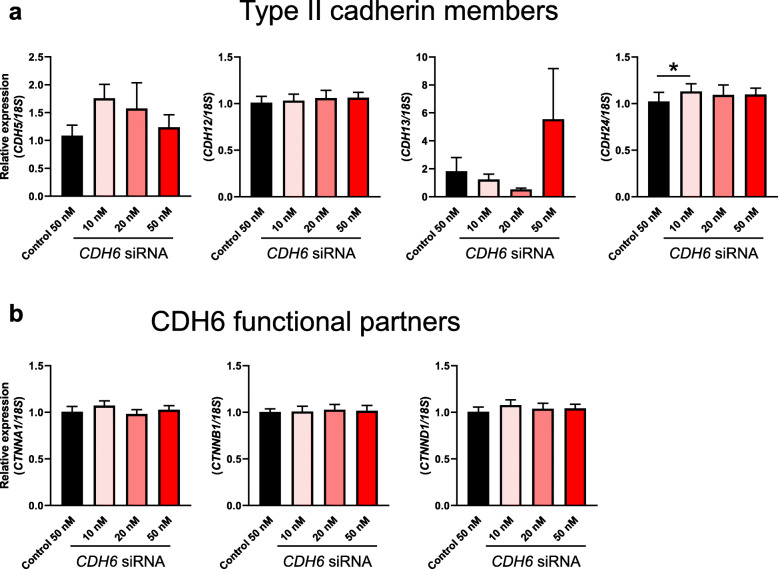
Methods: The expression and the localization of CDH6 in the human endometrium were determined by immunohistochemistry. Ishikawa cells were used to investigate the functional consequences of CDH6 knockdown on endometrial adhesive capacity to HTR8/SVneo (trophoblast cell line) spheroids in vitro. CDH6 knockdown was assessed by qPCR and immunoblotting. After CDH6 knockdown, the expression of type II cadherin family members and CDH6 functional partners were assessed by qPCR. Two-tailed unpaired student's t-test or one-way ANOVA as appropriate were used for statistical analysis with a significance threshold of P < 0.05.
Results: A significant reduction of CDH6 immunolocalization was recorded in the luminal and glandular epithelium of endometrium from women with infertility (P < 0.05) compared to fertile group respective cellular compartments in the mid-secretory phase. Functional analysis using Ishikawa cells demonstrated that knockdown of CDH6 (treated with 50 nM CDH6 siRNA) significantly reduced epithelial adhesive capacity (P < 0.05) to HTR8/SVneo spheroids compared to control and other type II cadherin family members likely failed to compensate for the loss of CDH6. The expression levels of CDH6 functional partners, catenin family members were not changed after CDH6 knockdown in Ishikawa cells.
Conclusion: Together, our data revealed that CDH6 was dysregulated in the endometrium from women with infertility and altered Ishikawa cell adhesive capacity. Our study supports a role for CDH6 in regulating endometrial adhesion and implantation.
Keywords: Adhesive molecules; CDH6; Embryo implantation; Endometrial epithelial cell; Endometrial receptivity; Trophoblast cell.
Conflict of interest statement
The authors report no competing interests.
Figures
Similar articles
-
Tripeptidyl peptidase I promotes human endometrial epithelial cell adhesive capacity implying a role in receptivity.Reprod Biol Endocrinol. 2020 Dec 14;18(1):124. doi: 10.1186/s12958-020-00682-0. Reprod Biol Endocrinol. 2020. PMID: 33317560 Free PMC article.
-
MAML1: a coregulator that alters endometrial epithelial cell adhesive capacity.Fertil Res Pract. 2021 Mar 27;7(1):8. doi: 10.1186/s40738-021-00100-y. Fertil Res Pract. 2021. PMID: 33773601 Free PMC article.
-
A novel "embryo-endometrial" adhesion model can potentially predict "receptive" or "non-receptive" endometrium.J Assist Reprod Genet. 2020 Jan;37(1):5-16. doi: 10.1007/s10815-019-01629-0. Epub 2019 Nov 27. J Assist Reprod Genet. 2020. PMID: 31776756 Free PMC article.
-
Implantation in the baboon: endometrial responses.Semin Reprod Endocrinol. 1999;17(3):257-65. doi: 10.1055/s-2007-1016233. Semin Reprod Endocrinol. 1999. PMID: 10797944 Review.
-
[The implantation receptive luteal phase of the endometrium. On the current status of molecular and cell biology research].Zentralbl Gynakol. 2001 Jun;123(6):319-27. doi: 10.1055/s-2001-16282. Zentralbl Gynakol. 2001. PMID: 11488159 Review. German.
Cited by
-
Tripeptidyl peptidase I promotes human endometrial epithelial cell adhesive capacity implying a role in receptivity.Reprod Biol Endocrinol. 2020 Dec 14;18(1):124. doi: 10.1186/s12958-020-00682-0. Reprod Biol Endocrinol. 2020. PMID: 33317560 Free PMC article.
-
MAML1: a coregulator that alters endometrial epithelial cell adhesive capacity.Fertil Res Pract. 2021 Mar 27;7(1):8. doi: 10.1186/s40738-021-00100-y. Fertil Res Pract. 2021. PMID: 33773601 Free PMC article.
-
Genome-Wide Association Studies, Runs of Homozygosity Analysis, and Copy Number Variation Detection to Identify Reproduction-Related Genes in Bama Xiang Pigs.Front Vet Sci. 2022 May 31;9:892815. doi: 10.3389/fvets.2022.892815. eCollection 2022. Front Vet Sci. 2022. PMID: 35711794 Free PMC article.
-
Towards an Improved Understanding of the Effects of Elevated Progesterone Levels on Human Endometrial Receptivity and Oocyte/Embryo Quality during Assisted Reproductive Technologies.Cells. 2022 Apr 21;11(9):1405. doi: 10.3390/cells11091405. Cells. 2022. PMID: 35563710 Free PMC article. Review.
-
A microphysiological model of human trophoblast invasion during implantation.Nat Commun. 2022 Mar 15;13(1):1252. doi: 10.1038/s41467-022-28663-4. Nat Commun. 2022. PMID: 35292627 Free PMC article.
References
-
- Boivin J, Bunting L, Collins JA, Nygren KG. International estimates of infertility prevalence and treatment-seeking: potential need and demand for infertility medical care. Hum Reprod. 2007;22:1506–1512. - PubMed
-
- Norwitz ER, Schust DJ, Fisher SJ. Implantation and the survival of early pregnancy. N Engl J Med. 2001;345:1400–1408. - PubMed
-
- Dimitriadis E, Nie G, Hannan NJ, Paiva P, Salamonsen LA. Local regulation of implantation at the human fetal-maternal interface. Int J Dev Biol. 2009;54:313–322. - PubMed
-
- Dimitriadis E, White C, Jones R, Salamonsen L. Cytokines, chemokines and growth factors in endometrium related to implantation. Hum Reprod Update. 2005;11:613–630. - PubMed
-
- Koot Y, Teklenburg G, Salker M, Brosens J, Macklon N. Molecular aspects of implantation failure. Biochim Biophys Acta. 1822;2012:1943–1950. - PubMed
MeSH terms
Substances
Grants and funding
LinkOut - more resources
Full Text Sources
Molecular Biology Databases
