. 2020 Jun 29;11(1):261.
doi: 10.1186/s13287-020-01778-5.

Cutaneous wound healing: canine allogeneic ASC therapy

Affiliations

Cutaneous wound healing: canine allogeneic ASC therapy

Nathaly Enciso et al. Stem Cell Res Ther. .

Abstract

Background: Wound healing is a complex biological process comprised of a series of sequential events aiming to repair injured tissue. Adult mesenchymal stem cells (MSCs) have been used in cellular therapy in preclinical animal studies; a promising source of MSCs is adipose tissue (AT). In this paper, we evaluated the clinical value and safety of the application of cultured allogenic MSCs from AT for acute and chronic skin wound healing in a canine model.

Methods: Twenty-four dogs of different breeds between 1 and 10 years of age with acute and chronic wounds were studied. Morphology of the wounded skin was monitored for changes over time via serial photographs and histopathological studies.

Results: The percentage of the wounds that exhibited contraction and re-epithelialization were significantly different between wounds treated with adipose mesenchymal stem cells (ASCs) and control wounds; this effect was observed in both acute and chronic conditions. At 90 days, re-epithelization of acute and chronic wounds reached more than 97%. Histopathological study revealed a reduction in inflammatory infiltrate and the presence of multiple hair follicles on day 7 after treatment with ASCs, promoting epidermal and dermal regeneration. To guarantee the safety of our treatment, we determined the serum levels of cytokine markers in our patients. ASC treatment upregulated granulocyte-macrophage colony stimulating factor (GM-CSF) at the gene level, which may contribute to the recruitment of cells that participate in skin repair to the site of injury.

Conclusions: The development of an allogenic ASC therapy to improve wound healing in a canine model could have a clinical impact in human treatment.

Keywords: ASC therapy; Adipose mesenchymal stem cells; Canine cutaneous wounds; Regenerative medicine.


Conflict of interest statement

The authors declare they have no competing interests.

Figures

Fig. 1
Fig. 1
Differentiation of canine adipose-derived mesenchymal stem cells (ASCs) into osteocyte, chondrocyte, and adipocyte lineages. a Representative photographs. ASCs from the 3rd passage showed spindle-shaped fibroblastic cell morphology (1). After 3 weeks, ASCs differentiated according to the conditioned medium added. Cells underwent von Kossa staining for osteogenic lineage (2), Alcian blue staining for chondrogenic lineage (3), and oil red O staining for adipogenic lineage (4) (bar length = 50 μm). b RT-qPCR analysis of transcription factors in the differentiation of ASCs. Data represent mean fold change values in genes ± SD compared to ASCs. ****A statistically significant difference of p < 0.0001 of each transcription factors representative of a specific lineage with respect to the genes of other lineages. n = 3 different experiments
Fig. 2
Fig. 2
Transmigration capacity. Quantification of cells migrating toward DMEM, DMEM + canine serum 10%, conditioned medium (CM), DMEM + GM CSF 200 ng/ml, and DMEM + GM CSF 100 ng/ml. Data represent the mean ± SD. *p < 0.05. ns, not significant. n = 3 different experiments
Fig. 3
Fig. 3
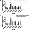
Cytokine concentration values in dogs using a Milliplex Canine Cytokine Panel. Canine serum levels of conventional and adipose-derived mesenchymal stem cell (ASC) treatment at 7 days (a) and 30 days (b) post-treatment. Values are the mean ± SD. **A statistically significant difference of p < 0.01. n = 4–6 for each experimental point
Fig. 4
Fig. 4
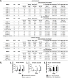
a Clinical evaluation. Wound contraction and regenerative area expressed in percentages in acute and chronic wounds. b Percentage of regenerated area of acute and chronic wounds after conventional and adipose-derived mesenchymal stem cell (ASC) treatment. Regenerated area for acute (1) and chronic (2) wounds treated with ASCs compared with conventional treatment at 7, 30, and 90 days. (3) Comparative studies between acute and chronic wounds treated with ASCs. Data values are the mean ± SD. ***A significant difference of p < 0.001. ****A significant difference of p < 0.0001. ns, no significant difference
Fig. 5
Fig. 5
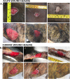
Re-epithelization of acute (a) and chronic (b) wounds is shown by representative photographs. Dogs with acute and chronic wounds treated with the conventional treatment (a (1–3), b (1–3)), and dogs treated with adipose-derived mesenchymal stem cells (ASCs) (a (4–6), b (4–6)) at day 0, 7, and 90
Fig. 6
Fig. 6
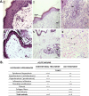
Histopathology of acute wound healing in control and ASC-treated skin wounds at 7 days post-treatment. a Representative photomicrographs demonstrate the histological characteristics of acute healing. H&E staining (bar length = 50 μm) (1 and 4), 200 μm (2 and 5), and 100 μm (3 and 6). (1–3) Control; (4–6) ASC-treated. H, hyperplasia; O, orthokeratosis; P, parakeratosis; Fb, fibrosis; MII, mononuclear inflammatory infiltrate; F, fibroblast; HF, hair follicles; C, collagen fibers. b Criteria for histological appearances in acute wounds with conventional treatment and ASC treatment. “-” and “+” = mild, “++” and “+++” = moderate, and “++++” = marked
Fig. 7
Fig. 7
Relative quantification of gene expression in acute skin wounds after 7 days of conventional and ASC treatments. Data represent mean fold change values in genes ± SD for both treatments as compared to normal skin. Data values are the mean ± SD. **A significant difference of p < 0.01. ***A significant difference of p < 0.001

References

    1. Hendijani F. Explant culture: An advantageous method for isolation of mesenchymal stem cells from human tissues. Cell proliferation. 2017;50:e12334. - PMC - PubMed
    1. Alstrup T, Eijken M, Bohn AB, Møller B, Damsgaard TE. Isolation of adipose tissue-derived stem cells: enzymatic digestion in combination with mechanical distortion to increase adipose tissue-derived stem cell yield from human aspirated fat. Curr Protoc Stem Cell Biol. 2019;48:e68. - PubMed
    1. Gentile P, Garcovich S. Concise review: adipose-derived stem cells (ASCs) and adipocyte-secreted exosomal microRNA (A-SE-miR) modulate cancer growth and promote wound repair. J Clin Med. 2019;8:855. - PMC - PubMed
    1. Gentile P, Calabrese C, De Angelis B, Pizzicannella J, Kothari A, Garcovich S. Impact of the different preparation methods to obtain human adipose-derived stromal vascular fraction cells (AD-SVFs) and human adipose-derived mesenchymal stem cells (AD-MSCs): enzymatic digestion versus mechanical centrifugation. Int J Mol Sci. 2019;20:5471. - PMC - PubMed
    1. Gentile P, Orlandi A, Scioli MG, Di Pasquali C, Bocchini I, Cervelli V. Concise review: adipose-derived stromal vascular fraction cells and platelet-rich plasma: basic and clinical implications for tissue engineering therapies in regenerative surgery. Stem Cells Transl Med. 2012;1:230–236. - PMC - PubMed

Publication types

LinkOut - more resources