. 2020 Aug 21;295(34):11949-11962.
doi: 10.1074/jbc.RA120.014555. Epub 2020 Jun 29.

PelX is a UDP- N-acetylglucosamine C4-epimerase involved in Pel polysaccharide-dependent biofilm formation

Affiliations

PelX is a UDP- N-acetylglucosamine C4-epimerase involved in Pel polysaccharide-dependent biofilm formation

Lindsey S Marmont et al. J Biol Chem. .

Abstract

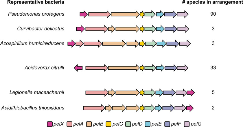
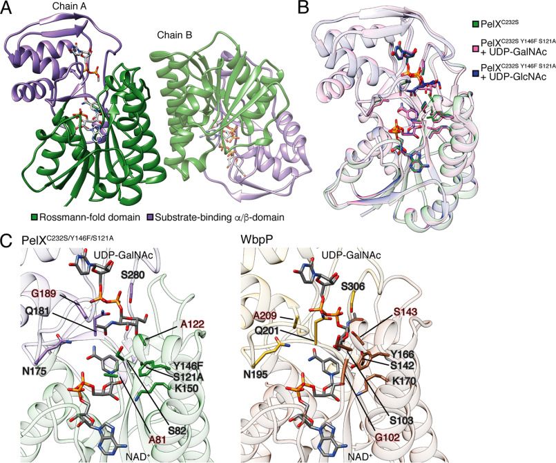
Pel is a GalNAc-rich bacterial polysaccharide that contributes to the structure and function of Pseudomonas aeruginosa biofilms. The pelABCDEFG operon is highly conserved among diverse bacterial species, and Pel may therefore be a widespread biofilm determinant. Previous annotation of pel gene clusters has helped us identify an additional gene, pelX, that is present adjacent to pelABCDEFG in >100 different bacterial species. The pelX gene is predicted to encode a member of the short-chain dehydrogenase/reductase (SDR) superfamily, but its potential role in Pel-dependent biofilm formation is unknown. Herein, we have used Pseudomonas protegens Pf-5 as a model to elucidate PelX function as Pseudomonas aeruginosa lacks a pelX homologue in its pel gene cluster. We found that P. protegens forms Pel-dependent biofilms; however, despite expression of pelX under these conditions, biofilm formation was unaffected in a ΔpelX strain. This observation led us to identify a pelX paralogue, PFL_5533, which we designate here PgnE, that appears to be functionally redundant to pelX In line with this, a ΔpelX ΔpgnE double mutant was substantially impaired in its ability to form Pel-dependent biofilms. To understand the molecular basis for this observation, we determined the structure of PelX to 2.1 Å resolution. The structure revealed that PelX resembles UDP-GlcNAc C4-epimerases. Using 1H NMR analysis, we show that PelX catalyzes the epimerization between UDP-GlcNAc and UDP-GalNAc. Our results indicate that Pel-dependent biofilm formation requires a UDP-GlcNAc C4-epimerase that generates the UDP-GalNAc precursors required by the Pel synthase machinery for polymer production.

Keywords: PelX; PgnE; Pseudomonas; Pseudomonas aeruginosa; Pseudomonas protegens; X-ray crystallography; bacterial adhesion; biofilm; enzyme; epimerase; microbiology; polysaccharide; short-chain dehydrogenase/reductase.


Conflict of interest statement

Conflict of interest—The authors declare that they have no conflicts of interest with the contents of this article.

Figures

Figure 1.
Figure 1.
A pelX homologous gene is found adjacent to pel biosynthetic gene clusters in three different arrangements. Each gene is shown as an arrow, where each arrow indicates the direction of transcription. The predicted function of each gene is indicated by its color as per the legend (bottom). One representative bacterial species is shown per gene arrangement. The total number of species identified with each arrangement is indicated (right). The full list of species with pelX-containing pel loci can be found in Dataset S1.
Figure 2.
Figure 2.
P. protegens forms Pel polysaccharide–dependent biofilms that are enhanced by the overexpression of the diguanylate cyclase, WspR. A and B, biofilm biomass determined using the crystal violet assay for (A) WT P. protegens Pf-5 and the corresponding deletion mutants assayed after 5 days of growth and (B) strains expressing pPSV39 (empty vector) or pPSV39::wspRR242A after 24 h of growth. Error bars indicate the S.E. of the mean of two (A) or three (B) independent trials performed in triplicate. Statistical significance was evaluated using one-way analysis of variance with Bonferroni correction. ***, p < 0.001; ****, p < 0.0001.
Figure 3.
Figure 3.
pelX or PFL_5533 is required for Pel polysaccharide–dependent biofilm formation by P. protegens. A, Western blotting of VSV-G–tagged proteins PelF, PelX, or PFL_5533 in the presence of native c-di-GMP levels (empty vector) or elevated c-di-GMP levels (WspRR242A). B, biofilm biomass as determined using the crystal violet assay after 6 h of static growth. Error bars indicate the S.E. of the mean of two independent trials performed in triplicate. Statistical significance was evaluated using one-way analysis of variance with Bonferroni correction; ***, p < 0.001; ****, p < 0.0001. C, dot blot of culture supernatants of the indicated strains probed using either horseradish peroxidase–conjugated WFL or the Pel antibody (α-Pel) (10, 27).
Figure 4.
Figure 4.
PelX is a Class 3 UDP-GlcNAc C4-epimerase. A and B, 1H NMR focused on the anomeric H-1 region of the spectra (red line) for (A) UDP-GlcNAc incubated without enzyme (top), UDP-GlcNAc with PelX (middle), and UDP-GalNAc with PelX (bottom) and (B) UDP-Glc incubated without enzyme (top), UDP-Glc with PelX (middle), and UDP-Gal with PelX (bottom). C, PelX catalyzes the epimerization of UDP-GlcNAc to UDP-GalNAc by inversion of the hydroxyl group at position C4. D, LC-MS/MS quantification of GlcNAc from cell extracts of the indicated strains. Error bars represent the S.E. of the mean of six independent biological replicates. Statistical significance was evaluated using unpaired t test. *, p < 0.05.
Figure 5.
Figure 5.
PelX is a member of the short-chain dehydrogenase/reductase superfamily of enzymes. Cartoon representations of PelXC232S. A, PelXC232S is displayed as found in the asymmetric unit with its N-terminal Rossmann fold domain shown in green, and its C-terminal substrate-binding α/β-domain in purple. B, overlay of PelXC232S/Y146F/S121A substrate complexes with the WT PelXC232S colored according to the legend in the figure. C, comparison of the active site of PelXC232S/Y146F/S121A–UDP-GalNAc complex, and WbpP–UDP-GalNAc complex (PDB ID: 1SB8). UDP-GalNAc and NADH (NAD+), and active site residues are shown in stick representation.

References

    1. Whitfield G. B., Marmont L. S., and Howell P. L. (2015) Enzymatic modifications of exopolysaccharides enhance bacterial persistence. Front. Microbiol. 6, 471 10.3389/fmicb.2015.00471 - DOI - PMC - PubMed
    1. Franklin M. J., Nivens D. E., Weadge J. T., and Howell P. L. (2011) Biosynthesis of the Pseudomonas aeruginosa extracellular polysaccharides, alginate, Pel, and Psl. Front. Microbiol. 2, 167 10.3389/fmicb.2011.00167 - DOI - PMC - PubMed
    1. Lee H. J., Chang H. Y., Venkatesan N., and Peng H. L. (2008) Identification of amino acid residues important for the phosphomannose isomerase activity of PslB in Pseudomonas aeruginosa PAO1. FEBS Lett. 582, 3479–3483 10.1016/j.febslet.2008.09.013 - DOI - PubMed
    1. Roychoudhury S., May T. B., Gill J. F., Singh S. K., Feingold D. S., and Chakrabarty A. M. (1989) Purification and characterization of guanosine diphospho-d-mannose dehydrogenase. A key enzyme in the biosynthesis of alginate by Pseudomonas aeruginosa. J. Biol. Chem. 264, 9380–9385 - PubMed
    1. Shinabarger D., Berry A., May T. B., Rothmel R., Fialho A., and Chakrabarty A. M. (1991) Purification and characterization of phosphomannose isomerase-guanosine diphospho-d-mannose pyrophosphorylase. A bifunctional enzyme in the alginate biosynthetic pathway of Pseudomonas aeruginosa. J. Biol. Chem. 266, 2080–2088 - PubMed

Publication types

MeSH terms

Substances

Supplementary concepts

Associated data

LinkOut - more resources