. 2020 Jul 14;117(28):16667-16677.
doi: 10.1073/pnas.2006620117. Epub 2020 Jun 29.

Rocks in the auxin stream: Wound-induced auxin accumulation and ERF115 expression synergistically drive stem cell regeneration

Affiliations

Rocks in the auxin stream: Wound-induced auxin accumulation and ERF115 expression synergistically drive stem cell regeneration

Balkan Canher et al. Proc Natl Acad Sci U S A. .

Erratum in

Abstract

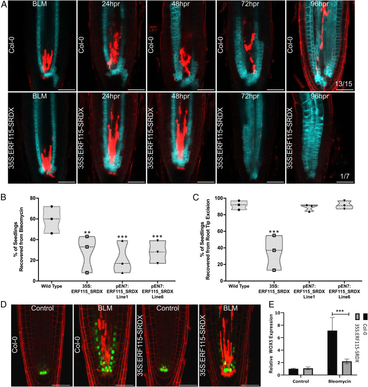
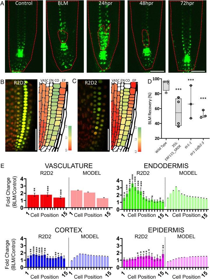
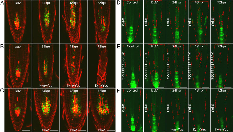
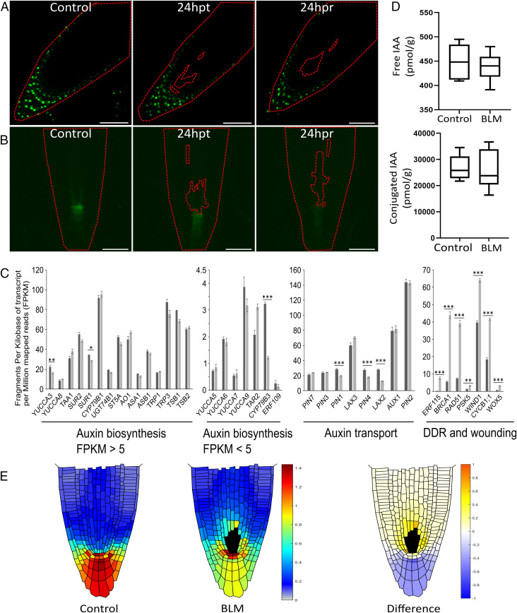
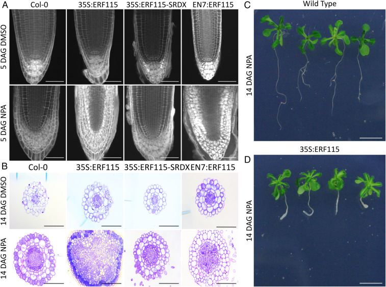
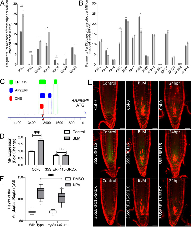
Plants are known for their outstanding capacity to recover from various wounds and injuries. However, it remains largely unknown how plants sense diverse forms of injury and canalize existing developmental processes into the execution of a correct regenerative response. Auxin, a cardinal plant hormone with morphogen-like properties, has been previously implicated in the recovery from diverse types of wounding and organ loss. Here, through a combination of cellular imaging and in silico modeling, we demonstrate that vascular stem cell death obstructs the polar auxin flux, much alike rocks in a stream, and causes it to accumulate in the endodermis. This in turn grants the endodermal cells the capacity to undergo periclinal cell division to repopulate the vascular stem cell pool. Replenishment of the vasculature by the endodermis depends on the transcription factor ERF115, a wound-inducible regulator of stem cell division. Although not the primary inducer, auxin is required to maintain ERF115 expression. Conversely, ERF115 sensitizes cells to auxin by activating ARF5/MONOPTEROS, an auxin-responsive transcription factor involved in the global auxin response, tissue patterning, and organ formation. Together, the wound-induced auxin accumulation and ERF115 expression grant the endodermal cells stem cell activity. Our work provides a mechanistic model for wound-induced stem cell regeneration in which ERF115 acts as a wound-inducible stem cell organizer that interprets wound-induced auxin maxima.

Keywords: Arabidopsis; ERF115; auxin; regeneration; stem cells.


Conflict of interest statement

The authors declare no competing interest.

Figures

Fig. 1.
Fig. 1.
(A and B) Time-lapse images of control and pSCR:CRE-GR 35S:loxp-tOCS-loxp-CFP seedlings in Col-0 or 35S:ERF115-SRDX background at indicated time points: 24 h of BLM treatment or 24 to 96 h of recovery on BLM-free medium. hpr, hours postrecovery. Propidium iodide was used to stain cell walls and damaged cells. The numbers in the lower right corner indicate the number of recovered seedlings among the total number of seedlings tracked by confocal imaging. (B and C) Percentage of seedlings recovered from BLM treatment (B) or root tip excision (C). **P < 0.01; ***P < 0.001. (D) WOX5_-GFP/NLS in the wild-type (Col-0) or 35S:ERF115-SRDX background under control conditions or following a 24-h treatment with BLM. Propidium iodide was used as a counterstain and to stain damaged cells. (E) qPCR data showing relative WOX5 expression in Col-0 and 35S:ERF115-SRDX before or after BLM treatment. ***P < 0.001. (Scale bars in A and D: 50 μm.)
Fig. 2.
Fig. 2.
(A) Confocal images of DR5:Venus-NLS under control conditions and during BLM recovery for indicated times. hpr, hours postrecovery. Images represent maximum intensity projections from 10 slices acquired at 1.5-µm intervals. Dashed red lines indicate the outline of the root and the region containing the dead cells, as inferred from propidium iodide staining (SI Appendix, Fig. S2A). (B and C) Confocal images of a R2D2 ratiometric auxin reporter line grown under control conditions (B) and after BLM treatment (C). Cartoons are representative images reconstructed from average R/G ratios calculated from 12 images. A higher red/green ratio corresponds to higher auxin levels. (Scale bars in A and B: 50 μm.) VASC, vasculature; EN, endodermis; CO, cortex; EP, epidermis. (D) BLM recovery percentages of wild-type, 35S:ERF115-SRDX, tir1-1, and tir1-1afb2-3 mutants. ***P < 0.001. (E) Side-by-side comparison of average fold changes in auxin concentrations as observed in vivo (R2D2) and predicted by the in silico model (MODEL). Error bars represent maximal errors calculated as described in Materials and Methods. Fold changes (BLM/control) in R/G ratios were obtained from R2D2 images according to the cell position in the respective tissues. Ratios from all cells within cell positions 1 to 5, 6 to 10, and 11 to 15 were averaged and plotted as three data points. *P < 0.05; **P < 0.01; ***P < 0.001.
Fig. 3.
Fig. 3.
Confocal images of pERF115:GFP-GUS (AC) and DR5:GFP (DF) seedlings in wild-type (A, B, C, D, and F) or 35S:ERF115-SRDX (E) background treated for 24 h with BLM and recovered on normal medium supplemented with DMSO (A, D, and E), with auxin biosynthesis inhibitors kynurenine and yucasin (B and F) or with 1 μM NAA (C). Red dashed lines in DF indicate the outline of the root and the region containing the dead stem cells as indicated by propidium iodide staining (SI Appendix, Fig. S3 DF). (Scale bars: 50 μm.)
Fig. 4.
Fig. 4.
(A and B) Confocal images of YUC9:VENUS (A) and TAA1:TAA1-GFP (B) seedlings before and after BLM treatment and indicated times of recovery. The red dashed line outlines the root and the location of dead cells as inferred by propidium iodide staining. hpt, hours posttreatment; hpr, hours postrecovery. (Scale bars: 50 μm.) (C) Transcriptional changes in auxin biosynthesis and transport genes and genes involved in the DNA damage response (DDR) and wounding, revealed by RNA-seq from BLM-treated root tips. *FDR < 0.05; **FDR < 0.01; ***FDR < 0.001. (D) Free and conjugated IAA concentrations before and after BLM treatment. (E) Auxin distributions obtained from an in silico model of polar auxin transport before and after BLM-induced stem cell death. The BLM picture represents the average obtained from simulation of 10 real-life cell death patterns. The plot represents the average fold changes in auxin concentrations per tissue against distance from the QC.
Fig. 5.
Fig. 5.
(A) Confocal images of pseudo-Schiff-stained Col-0, 35S:ERF115, 35S:ERF115-SRDX, and pEN7:ERF115 seedlings grown for 5 d on DMSO or 10 μM NPA. (B) Cross-sectional images of Col-0, 35S:ERF115, 35S:ERF115-SRDX, and EN7:ERF115 seedlings grown for 14 d on DMSO or 10 μM NPA. Sections correspond to the region 650 to 750 μm from the root tip. (C and D) Col-0 (C) and 35S:ERF115 (D) seedlings grown on 10 μM NPA for 14 d. (Scale bars: A and B, 100 μm; C and D, 1 cm.) DAG, days after germination.
Fig. 6.
Fig. 6.
(A and B) Fragments per kilobase of transcript per million reads (FPKM) of only differentially expressed AUX/IAA genes (A) and all ARF genes (B) obtained from RNA-seq transcriptome of BLM- vs. mock-treated seedlings. *P < 0.01; **P < 0.01; ***P < 0.001. (C) Putative AP2/ERF transcription factor binding regions (blue) according to O’Malley et al. (47), DNAse I hypersensitive site (DHS, red) according to Sulliven et al. (48), and putative ERF115-binding regions (green) according to Heyman et al. (46). (D) Fold changes in expression of MP with or without BLM treatment in Col-0 and 35S:ERF115-SRDX as indicated by qRT-PCR. (E) MP:MP-GFP reporter line in Col-0, 35S:ERF115, and 35S:ERF115-SRDX background before and after BLM treatment or 24 h post recovery (hpr). (Scale bars: 50 μm.) (F) Starch granule area quantification obtained from a segregating population of mpB4149 mutants. Homozygous mutant seedlings were not included in the analysis due to lack of a root.

References

    1. Aichinger E., Kornet N., Friedrich T., Laux T., Plant stem cell niches. Annu. Rev. Plant Biol. 63, 615–636 (2012). - PubMed
    1. Pi L. et al. ., Organizer-derived WOX5 signal maintains root columella stem cells through chromatin-mediated repression of CDF4 expression. Dev. Cell 33, 576–588 (2015). - PubMed
    1. Bennett T., Scheres B., Root development—two meristems for the price of one? Curr. Top. Dev. Biol. 91, 67–102 (2010). - PubMed
    1. Ha C. M., Jun J. H., Fletcher J. C., Shoot apical meristem form and function. Curr. Top. Dev. Biol. 91, 103–140 (2010). - PubMed
    1. Gutierrez L. et al. ., Phenotypic plasticity of adventitious rooting in Arabidopsis is controlled by complex regulation of AUXIN RESPONSE FACTOR transcripts and microRNA abundance. Plant Cell 21, 3119–3132 (2009). - PMC - PubMed

Publication types

MeSH terms