. 2020 Jul 14;117(28):16579-16586.
doi: 10.1073/pnas.2001637117. Epub 2020 Jun 29.

Yaravirus: A novel 80-nm virus infecting Acanthamoeba castellanii

Affiliations

Yaravirus: A novel 80-nm virus infecting Acanthamoeba castellanii

Paulo V M Boratto et al. Proc Natl Acad Sci U S A. .

Abstract

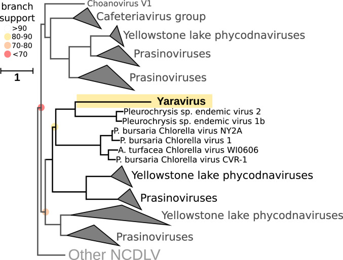
Here we report the discovery of Yaravirus, a lineage of amoebal virus with a puzzling origin and evolution. Yaravirus presents 80-nm-sized particles and a 44,924-bp dsDNA genome encoding for 74 predicted proteins. Yaravirus genome annotation showed that none of its genes matched with sequences of known organisms at the nucleotide level; at the amino acid level, six predicted proteins had distant matches in the nr database. Complimentary prediction of three-dimensional structures indicated possible function of 17 proteins in total. Furthermore, we were not able to retrieve viral genomes closely related to Yaravirus in 8,535 publicly available metagenomes spanning diverse habitats around the globe. The Yaravirus genome also contained six types of tRNAs that did not match commonly used codons. Proteomics revealed that Yaravirus particles contain 26 viral proteins, one of which potentially representing a divergent major capsid protein (MCP) with a predicted double jelly-roll domain. Structure-guided phylogeny of MCP suggests that Yaravirus groups together with the MCPs of Pleurochrysis endemic viruses. Yaravirus expands our knowledge of the diversity of DNA viruses. The phylogenetic distance between Yaravirus and all other viruses highlights our still preliminary assessment of the genomic diversity of eukaryotic viruses, reinforcing the need for the isolation of new viruses of protists.

Keywords: NCLDV; ORFan; Yaravirus; capsid; metagenomics.


Conflict of interest statement

The authors declare no competing interest.

Figures

Fig. 1.
Fig. 1.
Yaravirus particle and the beginning of the viral cycle. (A) Negative staining of an isolated Yaravirus virion. (Scale bar: 100 nm.) (B) Transmission electron microscopy (TEM) representing the beginning of the viral cycle, in which one particle is associated to the host cell membrane and the second one was already incorporated by the amoeba inside an endocytic vesicle. (Scale bar: 200 nm.) (C) Detailed image of an incorporated Yaravirus particle in the interior of an endocytic vesicle. (Scale bar: 100 nm.) (D) Viral uptake by the amoeba may occur individually but also in groups of particles, as observed in the micrograph. (Scale bar: 250 nm.) (E) The viral factory completely develops, occupying the nuclear region and recruiting mitochondria around it. Two different regions can be distinct: an electron-lucent region where the virions are assembled as empty shells and a second region formed by several electron-dense points where the genome is packaged inside the particles. (Scale bar: 500 nm.)
Fig. 2.
Fig. 2.
Yaravirus morphogenesis and release. (A) The virions are assembled by the addition of more than one layer of protein or membranous components around its structure. (Scale bar: 70 nm.) (B) The particles then start to migrate to the periphery of the cell, where there is the presence of several electron-dense points that function as morphogenetic structures to package the DNA inside the Yaravirus particles (regions inside dashed lines). (Scale bar: 1,000 nm.) (C) Detailed image of the morphogenetic regions where the DNA (red arrow) is incorporated inside the Yaravirus virion (black arrow). (Scale bar: 150 nm.) (D) Sometimes, the final step of viral replication is marked by the particles being packaged inside vesicle-like structures, suggesting a potential release by exocytosis. (Scale bar: 500 nm.) (E) Most of the particles, however, are released by cellular lysis and have a high affinity to the membranes of cellular debris. (Scale bar: 150 nm.) (F) Graph comparing concomitantly the decrease of host cell numbers (red bars) with the increase of Yaravirus genome during the infection (black line). Replication of viral genome was measured by qPCR and calculated by delta-delta Ct.
Fig. 3.
Fig. 3.
Yaravirus genome features. (A) Circular representation of Yaravirus genome highlighting the predicted ORFs (arrows). Red arrows represent ORFs predicted by analyses of similarity of amino acid sequences with information regarding their best hits. Black dots indicate ORFs encoding predicted proteins whose functions were suggested by HHPred (structural analyses). Yellow stars indicate proteins found in virion proteomics. (B) The percentage of ORFan genes among the complete genome of different viruses of amoeba is represented by the graph with red scale bars. (C) The graph with greenish scale bars represents the absolute number of genes with homologs in databases (non-ORFan genes) for each of the same amoebal viruses previously analyzed. (D) All of the six Yaravirus predicted tRNAs, as well as their corresponding sequences, are pictured with information about their anticodon (in parentheses), their nucleotide length, the percentage of GC content, and the position in the intergenic regions of genes 29 and 30. Regarding the asterisk, note that the number of ORFan/non-ORFan genes represented in C and D do not take into account the structural annotation made by the HHpred servers, as for most of the amoebal viruses represented in the graphs.
Fig. 4.
Fig. 4.
Phylogenetic position of Yaravirus and related viral sequences in the Mimiviridae based on the viral ATPase (NCVOG0249). The Yaravirus ATPase is highlighted in yellow. Branch support is indicated as colored circles for support values of 90 or below. The tree is rooted at the Poxviruses. (Scale bar: substitutions per site.) GC content of viral genomes and contigs containing NCVOG0249 is shown together with the average GC content of collapsed clades. In addition, environmental origin and assembly sizes of Yaravirus and related viral contigs and genomes are shown.
Fig. 5.
Fig. 5.
Structure-guided phylogeny based on hmmsearch employed to identify the Yaravirus MCP in 235 NCDLV reference genomes using specific hidden Markov models. The alignments were made with Expresso in the software T-Coffee, using PDB structures. After, the phylogenetic trees were built using IQ-tree (v1.6.12; ref. 61) with LG+F+R8 based on the built-in model select feature (62) and 1,000 ultrafast bootstrap replicates (63). The Yaravirus MCP is highlighted in yellow. Branch support is indicated as colored circles for support values of 90 or below. The tree is unrooted. (Scale bar: substitutions per site.)

References

    1. Guglielmini J., Woo A. C., Krupovic M., Forterre P., Gaia M., Diversification of giant and large eukaryotic dsDNA viruses predated the origin of modern eukaryotes. Proc. Natl. Acad. Sci. U.S.A. 116, 19585–19592 (2019). - PMC - PubMed
    1. Koonin E. V., Yutin N., Multiple evolutionary origins of giant viruses. F1000 Res. 7, 1840 (2018). - PMC - PubMed
    1. Colson P. et al. ., Ancestrality and mosaicism of giant viruses supporting the definition of the fourth TRUC of microbes. Front. Microbiol. 9, 2668 (2018). - PMC - PubMed
    1. Colson P., Ominami Y., Hisada A., La Scola B., Raoult D., Giant mimiviruses escape many canonical criteria of the virus definition. Clin. Microbiol. Infect. 25, 147–154 (2019). - PubMed
    1. Abrahão J. et al. ., Tailed giant Tupanvirus possesses the most complete translational apparatus of the known virosphere. Nat. Commun. 9, 749 (2018). - PMC - PubMed

Publication types

Associated data