. 2020 Jun 29;12(13):12943-12959.
doi: 10.18632/aging.103378. Epub 2020 Jun 29.

Nrf2 inhibits ferroptosis and protects against acute lung injury due to intestinal ischemia reperfusion via regulating SLC7A11 and HO-1

Affiliations

Nrf2 inhibits ferroptosis and protects against acute lung injury due to intestinal ischemia reperfusion via regulating SLC7A11 and HO-1

Hui Dong et al. Aging (Albany NY). .

Erratum in

Abstract

Acute lung injury (ALI) is a syndrome associated with a high mortality rate. Nrf2 is a key regulator of intracellular oxidation homeostasis that plays a pivotal role in controlling lipid peroxidation, which is closely related to the process of ferroptosis. However, the intrinsic effect of Nrf2 on ferroptosis remains to be investigated in ALI. We found that MDA expression increased while GSH and GPX4 decreased in ALI models. Furthermore, the characteristic mitochondrial morphological changes of ferroptosis appear in type II alveolar epithelial cells in IIR models. Additional pre-treatment of Fe and Ferrostatin-1 in ALI significantly aggravated or ameliorated the pathological injuries of lung tissue, pulmonary edema, lipid peroxidation, as well as promoted or prevented cell death, respectively. Knocking down Nrf2 notably decreased the expression of SLC7A11 and HO-1. Interference with SLC7A11 markedly increased Nrf2-HO-1 and dramatically attenuated cell death in OGD/R models. These findings indicate that ferroptosis can be inhibited by Nrf2 through regulating SLC7A11 and HO-1, which may provide a potential therapeutic strategy for IIR-ALI.

Keywords: acute lung injury; ferroptosis; heme oxygenase-1; solute carrier family 7 member 11; uclear factor erythroid 2 related factor2.


Conflict of interest statement

CONFLICTS OF INTEREST: The authors declare no competing financial interests.

Figures

Figure 1
Figure 1
IIR induces ferroptosis in type II alveolar epithelial cells. (A) The degree of pulmonary oedema continued to increase with the extension of reperfusion time. (B) Representative transmission electron micrographs of the ultrastructure of lung tissues. Scale bars: 1 μm. (C) HE staining of the lung tissues of mice following IIR, IIR + Fe, and IIR + Fer-1. Scale bars: 200 μm. (D) The lung pathological damage score showed an addition and reduction after Fe and Fer-1 administration, respectively. (E) The wet to dry ratio of the lung tissue shown in each group. (F) The level of lipid peroxide MDA in each group. (G) The GSH level in each group. The error bars represent the standard error from three replicates. Data are presented as the mean ± SEM. *P < 0.05; **P < 0.01; ***P < 0.001 between the groups; *compared with the sham group; # compared with the IIR group.
Figure 2
Figure 2
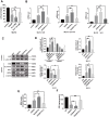
Nrf2 regulates SLC7A11 and HO-1 to inhibit ferroptosis and protect against IIR-ALI. (A) Western blot analysis of Nrf2, HO-1, SLC7A11, and GPX4 expression in the lung tissues of each group. (B) The representative quantification of these proteins. (C) Relative mRNA expression of Nrf2, SLC7A11, and HO-1 in each group. (D) Representative transmission electron micrographs of the ultrastructure of the lung tissues. Scale bars: 1 μm. (E) Representative HE-stained lung sections. Morphology was examined using light microscopy. Scale bars: 200 μm. (F) Pathological scores were assigned by an experienced pathologist. (G) Western blot analysis of Nrf2, SLC7A11, and HO-1 expression in the lung tissues of each group. (H) The representative quantification of these proteins. (I) Relative mRNA expression of SLC7A11 and HO-1 in each group. (J) Western blot analysis of GPX4 expression in each group. (K) The representative quantification of GPX4 protein. (L) The lipid peroxide MDA level in each group. (M) The GSH level in each group. The error bars represent the standard error from three replicates. Data are presented as the mean ± SEM. *P < 0.05; **P < 0.01;***P < 0.001. # compared with the IIR group in WT mice.
Figure 3
Figure 3
OGD/R induces ferroptosis in pulmonary epithelial cells and increases the level of Nrf2 /SLC7A11/HO-1 expression during ferroptosis. (A) The cells surviving after OGD (8 h)/R (12 h), while the administration of Fe (3.3M) (800 μg/mL)/Fer-1 (0.1 μM) can respectively increase or decrease the ratio. (B) Relative mRNA expression of Nrf2, HO-1, and SLC7A11 in MLE12. (C) Western blotting of Nrf2, HO-1, GPX4, and SLC7A11 protein expression in each group. (D) The representative quantification of these proteins. (E) The level of lipid peroxide MDA in each group. (F) The GSH level in each group. The error bars represent the standard error from three replicates. Data are presented as the mean ± SEM. *P < 0.05; **P < 0.01; ***P < 0.001; *compared with control; # compared with OGD/R.
Figure 4
Figure 4
Nrf2 activation contributes to ferroptosis resistance. The cells were transfected with lentiviruses (A and F). Western blot analysis of the level of Nrf2, HO-1, and SLC7A11 protein expression in each group and the representative quantification of these proteins. (B and G) Cell viability was determined by a CCK-8 assay (n = 3). (C and H) Western blot analysis of the level of GPX4 protein expression in each group and the representative quantification of GPX4. (D and I) The level of lipid peroxide MDA in each group. (E and J) The level of GSH in each group. The error bars represent the standard error from three replicates. Data are presented as the mean ± SEM. *P < 0.05; **P < 0.01; ***P < 0.001.
Figure 5
Figure 5
Low levels of SLC7A11 alleviate cell death by upregulating Nrf2-HO-1, whereas SLC7A11 overexpression (OE-SLC7A11) enhanced cell death. (A and C) Western blot analysis of the Nrf2, HO-1, and SLC7A11 in each group and the representative quantification of these proteins in MLE12 cells. (B and D) Cell viability was determined using a CCK-8 assay (n = 3). The error bars represent the standard error from three replicates. Data are presented as the mean ± SEM. *P < 0.05; **P < 0.01; ***P < 0.001.

References

    1. Zhang F, Li ZL, Xu XM, Hu Y, Yao JH, Xu W, Jing HR, Wang S, Ning SL, Tian XF. Protective effects of icariin-mediated SIRT1/FOXO3 signaling pathway on intestinal ischemia/reperfusion-induced acute lung injury. Mol Med Rep. 2015; 11:269–76. 10.3892/mmr.2014.2679 - DOI - PubMed
    1. Meng QT, Chen R, Chen C, Su K, Li W, Tang LH, Liu HM, Xue R, Sun Q, Leng Y, Hou JB, Wu Y, Xia ZY. Transcription factors Nrf2 and NF-κB contribute to inflammation and apoptosis induced by intestinal ischemia-reperfusion in mice. Int J Mol Med. 2017; 40:1731–40. 10.3892/ijmm.2017.3170 - DOI - PMC - PubMed
    1. Stallion A, Kou TD, Latifi SQ, Miller KA, Dahms BB, Dudgeon DL, Levine AD. Ischemia/reperfusion: a clinically relevant model of intestinal injury yielding systemic inflammation. J Pediatr Surg. 2005; 40:470–77. 10.1016/j.jpedsurg.2004.11.045 - DOI - PubMed
    1. Mura M, Andrade CF, Han B, Seth R, Zhang Y, Bai XH, Waddell TK, Hwang D, Keshavjee S, Liu M. Intestinal ischemia-reperfusion-induced acute lung injury and oncotic cell death in multiple organs. Shock. 2007; 28:227–38. 10.1097/01.shk.0000278497.47041.e3 - DOI - PubMed
    1. Cavriani G, Oliveira-Filho RM, Trezena AG, da Silva ZL, Domingos HV, de Arruda MJ, Jancar S, Tavares de Lima W. Lung microvascular permeability and neutrophil recruitment are differently regulated by nitric oxide in a rat model of intestinal ischemia-reperfusion. Eur J Pharmacol. 2004; 494:241–49. 10.1016/j.ejphar.2004.04.048 - DOI - PubMed

Publication types

MeSH terms

LinkOut - more resources