. 2020 Jun 29;10(1):10590.
doi: 10.1038/s41598-020-67419-2.

Analyzing microglial-associated Aβ in Alzheimer's disease transgenic mice with a novel mid-domain Aβ-antibody

Affiliations

Analyzing microglial-associated Aβ in Alzheimer's disease transgenic mice with a novel mid-domain Aβ-antibody

Kristi Henjum et al. Sci Rep. .

Abstract

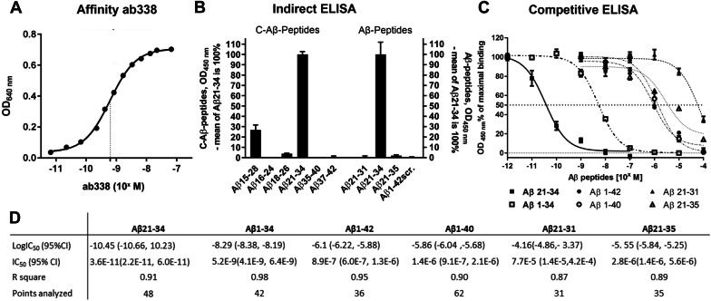
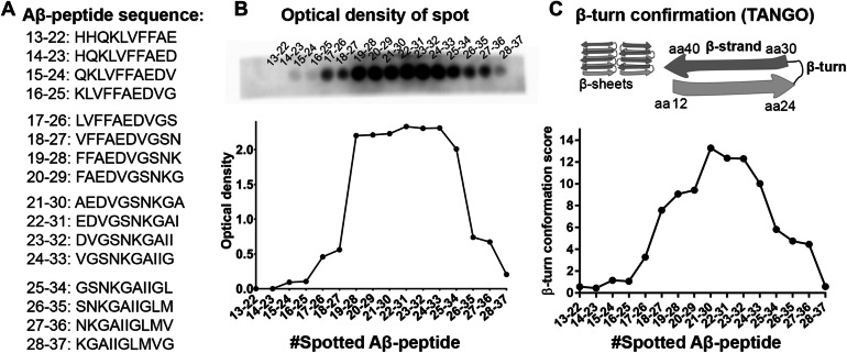
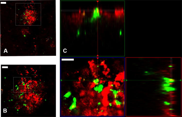
The mechanisms of amyloid-β (Aβ)-degradation and clearance in Alzheimer's disease (AD) pathogenesis have been relatively little studied. Short Aβ-fragments form by enzymatic cleavage and alternate amyloid-beta precursor protein (APP)-processing. Here we characterized a novel polyclonal Aβ-antibody raised against an Aβ mid-domain and used it to investigate microglial Aβ-uptake in situ by microscopy at the light- and ultrastructural levels. The rabbit Aβ-mid-domain antibody (ab338), raised against the mid-domain amino acids 21-34 (Aβ21-34), was characterized with biochemical and histological techniques. To identify the epitope in Aβ recognized by ab338, solid phase and solution binding data were compared with peptide folding scores as calculated with the Tango software. The ab338 antibody displayed high average affinity (KD: 6.2 × 10⁻¹⁰ M) and showed preference for C-terminal truncated Aβ-peptides ending at amino acid 34 and Aβ-mid domain peptides with high scores of β-turn structure. In transgenic APP-mouse brain, ab338 labelled amyloid plaques and detected Aβ-fragments in microglia at the ultra- and light microscopic levels. This reinforces a role of microglia/macrophages in Aβ-clearance in vivo. The ab338 antibody might be a valuable tool to study Aβ-clearance by microglial uptake and Aβ-mid-domain peptides generated by enzymatic degradation and alternate production.


Conflict of interest statement

The authors declare no competing interests.

Figures

Figure 1
Figure 1
Ab338 binding to Aβ depends on a mid-domain region in Aβ and amino acid 34. (A) Average affinity of the ab338 antibody to immobilized KLH-Cys-Aβ21–34 peptide as measured by optic density (OD) absorbance with points presented as mean ± standard error of the mean (SEM; n = 3). The data are presented as an adapted sigmoid curve and the dotted line indicates the equillibrum dissociation constant, KD. (B) Indirect ELISA displaying binding of ab338 to various immobilized Aβ-peptides, some of which harbor a N-terminal cystein (C-Aβ-peptides). Binding are presented relative to Aβ21–34 as mean ± SEM after vehicle coat background subtraction. Data are from two independent experiments (n = 6 in two experiments, total n = 12). (C) Competitive ELISA with various Aβ-peptides competing in solution for binding to ab338 binding with the KLH-Cys-Aβ21–34 plate coat presented with the sigmoid adapted curves (n ≥ 6, from ≥ 2 independent experiments). (D) Half inhibitory concentration (IC50) of various Aβ-peptides competing for ab338 binding with coat in competitive ELISA.
Figure 2
Figure 2
Binding of antibody ab338 to spot-synthesized Aβ-peptides relates to β-turn score in Aβ. (A) The amino acid sequences of the spot-synthesized Aβ-peptides with amino acid numbers in the Aβ-domain. (B) Antibody ab338 binding to Aβ-sequences spot-synthesized to membrane (top) and a graphical illustration of the optical density as a measure of ab338 preference for the spot-synthesized peptides. A high signal from Aβ-peptide sequences starting from amino acid 19–28 and ending at 24–33 (25–34) corresponded to a (C) high β-turn score of Aβ-peptides calculated by Tango. Illustration of Aβ-fibrils with β-sheets of β-strands and β-turns formed by Aβ-peptide. The drawing is based on the Aβ40-fibril structural model of sequences and are presented as suggested by Petkova et al. (2002), PNAS.
Figure 3
Figure 3
Microglial filopodia with Aβ in tgArcSwe mouse brain. (A) The microglia surround the amyloid plaques as illustrated here by microglia labelling (tomato lectin; red label) and an amyloid plaque (ab338; green label) in a 12 months-old transgenic tgArcSwe mouse. Nuclei are labelled by DAPI (blue label). (B) In the rim, Aβ-staining (ab338) is located in ramified processes of microglia. (C) Z-stacking of a microglia shows Aβ-staining (ab338) within filopodia as illustrated by an ortho-image and (D) the fluorescence profile. Images are obtained by confocal microscopy at 63 × magnification with increasing digital zoom. The scale bars measure 10, 5 and 5 µM in images (AC) respectively.
Figure 4
Figure 4
Aβ is located in microglial lysosomes. (A) TgSwe brain section showing an amyloid plaque surrounded by microglia with double staining using antibodies ab338 (red) and microglia by lysosomal marker CD68 (green), and (B) depicted with increased digital zoom. (C) Ortho image illustrating CD68- and ab338-staining being colocalized (yellow spots). The scale bars measure 20 µM, 10 µM and 5 µM and the gamma is set at 1.38/1.50, 1.42/1.58 and 1.42/1.42 (green/red channels) in (AC) respectively. The images were obtained with a confocal microscope.
Figure 5
Figure 5
Ultrastructural illustration of a microglia located close to Aβ-immunoreactive fibrillar material in tgArcSwe mouse brain. (A) Transmission electron microscopy of tgArcSwe mouse brain showing a microglia (M) and a blood vessel (V). (B) At higher magnification, the ultrastructural microglial morphology of the microglial cell is appearent with Aβ-fibrils visualized by ab338 immuno-gold labelling in the rim of and in the microglia. (C) Focusing on the microglia that are attracted towards the amyloid fibrils and ab388 also label within the microglia suggesting Aβ-uptake. For structural information, fibrillar labelling with ab388 is illustrated by arrows (B,C). The scale bars measure 5 µm (A), 0.5 µm (B) and 0.2 µm (C).

References

    1. Barker WW, et al. Relative frequencies of Alzheimer disease, Lewy body, vascular and frontotemporal dementia, and hippocampal sclerosis in the State of Florida Brain Bank. Alzheimer Dis. Assoc. Disord. 2002;16:203–212. doi: 10.1097/00002093-200210000-00001. - DOI - PubMed
    1. Duyckaerts C, Delatour B, Potier MC. Classification and basic pathology of Alzheimer disease. Acta Neuropathol. 2009;118:5–36. doi: 10.1007/s00401-009-0532-1. - DOI - PubMed
    1. Haass C, Selkoe DJ. Soluble protein oligomers in neurodegeneration: Lessons from the Alzheimer's amyloid beta-peptide. Nat. Rev. Mol. Cell Biol. 2007;8:101–112. doi: 10.1038/nrm2101. - DOI - PubMed
    1. Hardy J, Selkoe DJ. The amyloid hypothesis of Alzheimer's disease: Progress and problems on the road to therapeutics. Science. 2002;297:353–356. doi: 10.1126/science.1072994. - DOI - PubMed
    1. Heneka MT, et al. Neuroinflammation in Alzheimer's disease. Lancet Neurol. 2015;14:388–405. doi: 10.1016/s1474-4422(15)70016-5. - DOI - PMC - PubMed

Publication types

MeSH terms

Substances

LinkOut - more resources