. 2020 Jun 29;10(1):10552.
doi: 10.1038/s41598-020-67440-5.

Effects of topical corticosteroids and lidocaine on Borrelia burgdorferi sensu lato in mouse skin: potential impact to human clinical trials

Affiliations

Effects of topical corticosteroids and lidocaine on Borrelia burgdorferi sensu lato in mouse skin: potential impact to human clinical trials

Bastien Lefeuvre et al. Sci Rep. .

Abstract

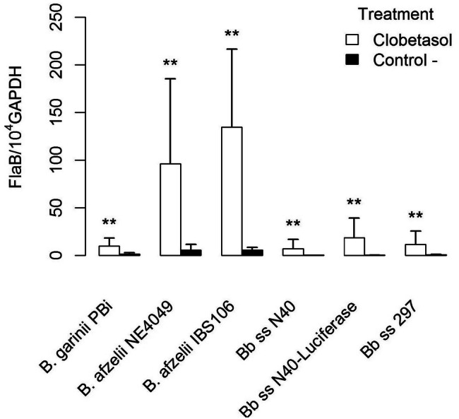
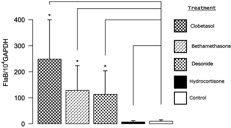
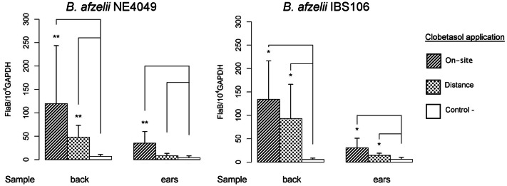
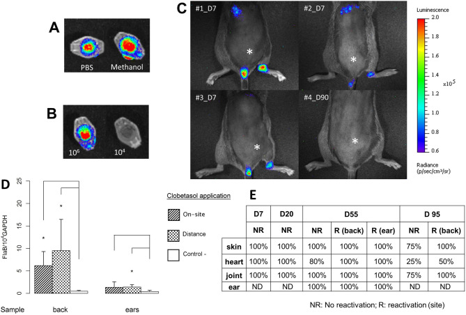
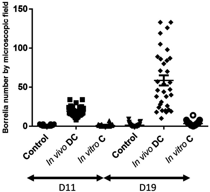
Lyme borreliosis is the most prevalent vector-borne disease in northern hemisphere. Borrelia burgdorferi sensu lato spirochetes are transmitted by Ixodes species ticks. During a blood meal, these spirochetes are inoculated into the skin where they multiply and often spread to various target organs: disseminated skin sites, the central nervous system, the heart and large joints. The usual diagnosis of this disease relies on serological tests. However, in patients presenting persistent clinical manifestations, this indirect diagnosis is not capable of detecting an active infection. If the serological tests are positive, it only proves that exposure of an individual to Lyme spirochetes had occurred. Although culture and quantitative PCR detect active infection, currently used tests are not sensitive enough for wide-ranging applications. Animal models have shown that B. burgdorferi persists in the skin. We present here our targeted proteomics results using infected mouse skin biopsies that facilitate detection of this pathogen. We have employed several novel approaches in this study. First, the effect of lidocaine, a local anesthetic used for human skin biopsy, on B. burgdorferi presence was measured. We further determined the impact of topical corticosteroids to reactivate Borrelia locally in the skin. This local immunosuppressive compound helps follow-up detection of spirochetes by proteomic analysis of Borrelia present in the skin. This approach could be developed as a novel diagnostic test for active Lyme borreliosis in patients presenting disseminated persistent infection. Although our results using topical corticosteroids in mice are highly promising for recovery of spirochetes, further optimization will be needed to translate this strategy for diagnosis of Lyme disease in patients.


Conflict of interest statement

The authors declare no competing interests.

Figures

Figure 1
Figure 1
Comparison of reactivation of several Borrelia burdgdorferi sensu lato strains as detected by qPCR in the skin at the site of inoculation (lower back) after occurrence of disseminated infection (40–50 days post inoculation). Different groups were compared by a Mann–Whitney test and p values for each strain from left to right are: 0.003, 0.002, 0.008, 0.006, 0.002, and 0.001.
Figure 2
Figure 2
Determination of B. afzelii NE4049 reactivation at day 50 by qPCR, at the site of inoculation after application of topical corticoids with different levels of anti-inflammatory activities: clobetasol propionate 0.05% (very high); bethamethasone valerate 0.1% (high); desonide 0.1% (high); hydrocortisone 0.5% (low); control (non-reactivated infected skin). Different groups were compared by a Mann–Whitney test and p values relative to control from left to right are: 0.02, 0.02, 0.02, and 0.4.
Figure 3
Figure 3
Quantification of B. afzelii NE4049 and IBS 106 by real-time qPCR after application of clobetasol on the back or on the ears of mice 40–50 days after Borrelia inoculation in the lower back of mice. Control (−) depicts infected skin collected from the back without reactivation. Different groups were compared by a Mann–Whitney test and p values relative to control from left to right for NE4049 are: 0.002, 0.002, 0.002, and 0.08, and for IBS 106 are: 0.01, 0.01, 0.02, and 0.01.
Figure 4
Figure 4
Live-imaging of luciferase-expressing B. burgdorferi strain N40D10/E9. In vitro assay was first performed to select the most suitable solvent to be used. (A) d-Luciferin substrate was diluted either in PBS or in methanol in Eppendorf tube to select the best solvent to detect bioluminescence. (B) Examination of two luciferase-expressing N40 concentrations added to the Eppendorf tubes, 104 and 106 spirochetes, with the substrate. In vivo assay: (C) Measurement of luciferase-expressing B. burgdorferi ss N40 strain injected in C3H/HeN mice at 7 days post-infection (natural peak of Borrelia multiplication-3 mice were selected) or 90 days (clobetasol reactivation—mouse#4) post-infection. The intradermal injection site of luciferase-expressing Borrelia is marked by an asterisk. (D) Quantification of luciferase-expressing N40 strain in the skin of C3H/HeN mice at day 55: the site of Borrelia inoculation was always at the lower back of mice, but the clobetasol reactivation was either “on site” of inoculation or “at distance”. Infected but non-reactivated mice were included as controls. The skin was collected either from the back (site of inoculation) or from the ear (distant skin from the inoculation site). Different groups were compared by a Mann–Whitney test and p values relative to control mice from left to right for back are: 0.01 and 0.01, and for ear are: 0.2 and 0.01. (E) The study was completed by culture of other tissues at different time points: heart, right joint, skin at the site of inoculation (back) and skin at a distant site (ear). The clobetasol application site is marked by an ‘R’ for reactivated mice (Day 55 and 95) or ‘NR’ for infected skin but ‘not reactivated’. “%” positive in this table was calculated using the number of mouse organ from which Borrelia could be recovered by culture with respect to the total number of samples cultured and examined.
Figure 5
Figure 5
The effect of clobetasol (DC) application in vivo or incubation of B. afzelii NE4049-infected skin with cortisone (C) in vitro at day 40 of inoculation on the lower back of mice. Infected skins (3 biopsy punches per mouse) were incubated in BSK-H complete medium at 33 °C for 11 and 19 days and: cultured directly for control (infected but non-reactivated mouse), reactivated in vivo by skin application of clobetasol (DC) and cultured, or cultured directly with cortisone included in culture medium. Borreliae were counted by dark-field microscopy using a Petroff-Hausser chamber at two time points.

References

    1. Steere A, et al. Lyme borreliosis. Nat Rev Dis Prim. 2016;2:1–13. - PMC - PubMed
    1. Stanek G, Wormser G, Gray J, Strle F. Lyme borreliosis. Lancet. 2012;379:461–473. doi: 10.1016/S0140-6736(11)60103-7. - DOI - PubMed
    1. Stanek G, et al. Lyme borreliosis: clinical case definitions for diagnosis and management in Europe. Clin Microbiol Infect. 2011;17:69–79. doi: 10.1111/j.1469-0691.2010.03175.x. - DOI - PubMed
    1. Borchers A, Keen C, Huntley A, Gershwin M. Lyme disease: a rigorous review of diagnostic criteria and treatment. J Autoimmun. 2015;57:82–115. doi: 10.1016/j.jaut.2014.09.004. - DOI - PubMed
    1. Wormser GP, et al. Practice guidelines for the treatment of lyme disease. Clin Infect Dis. 2000;31:S1–S14. doi: 10.1086/314053. - DOI - PubMed

Publication types

MeSH terms