. 2020 Oct;100(10):1288-1299.
doi: 10.1038/s41374-020-0455-y. Epub 2020 Jun 29.

Multiclass cancer classification in fresh frozen and formalin-fixed paraffin-embedded tissue by DigiWest multiplex protein analysis

Affiliations

Multiclass cancer classification in fresh frozen and formalin-fixed paraffin-embedded tissue by DigiWest multiplex protein analysis

Teresa Bockmayr et al. Lab Invest. 2020 Oct.

Abstract

Histomorphology and immunohistochemistry are the most common ways of cancer classification in routine cancer diagnostics, but often reach their limits in determining the organ origin in metastasis. These cancers of unknown primary, which are mostly adenocarcinomas or squamous cell carcinomas, therefore require more sophisticated methodologies of classification. Here, we report a multiplex protein profiling-based approach for the classification of fresh frozen and formalin-fixed paraffin-embedded (FFPE) cancer tissue samples using the digital western blot technique DigiWest. A DigiWest-compatible FFPE extraction protocol was developed, and a total of 634 antibodies were tested in an initial set of 16 FFPE samples covering tumors from different origins. Of the 303 detected antibodies, 102 yielded significant correlation of signals in 25 pairs of fresh frozen and FFPE primary tumor samples, including head and neck squamous cell carcinomas (HNSC), lung squamous cell carcinomas (LUSC), lung adenocarcinomas (LUAD), colorectal adenocarcinomas (COAD), and pancreatic adenocarcinomas (PAAD). For this signature of 102 analytes (covering 88 total proteins and 14 phosphoproteins), a support vector machine (SVM) algorithm was developed. This allowed for the classification of the tissue of origin for all five tumor types studied here with high overall accuracies in both fresh frozen (90.4%) and FFPE (77.6%) samples. In addition, the SVM classifier reached an overall accuracy of 88% in an independent validation cohort of 25 FFPE tumor samples. Our results indicate that DigiWest-based protein profiling represents a valuable method for cancer classification, yielding conclusive and decisive data not only from fresh frozen specimens but also FFPE samples, thus making this approach attractive for routine clinical applications.


Conflict of interest statement

GE, JS, AA, and CS are employees of NMI TT Pharmaservices, a company offering DigiWest service studies. All other authors declare no conflicts of interest.

Figures

Fig. 1
Fig. 1. DigiWest protein profiling in fresh frozen and FFPE tissue.
a DigiWest data displayed for four antibodies (Cytokeratin 5, p53, β-Catenin-phospho S675, HSP 27-phospho S78) in different tumor samples (fresh frozen in blue and FFPE tissue in red) with signal intensity (RFU: relative fluorescence units) plotted against molecular weight. b Pearson correlation coefficients (R) were computed for each antibody between all signals detected in both fresh frozen and FFPE samples. p values were corrected for multiple testing with the Benjamini–Hochberg method (p-BH), using a significance level of 0.05. The corresponding volcano plot shows the p values (−log10 transformed) plotted against correlation coefficients (R) (n: number of antibodies with significant correlation (p-BH < 0.05), colored in orange; m: number of antibodies with p-BH > 0.05, colored in blue). c Relative signals (log2) detected by DigiWest for three antibodies (Cytokeratin 5, c-Myc, Caspase 6) in 25 tumor samples, in both fresh frozen and FFPE samples. COAD colorectal adenocarcinomas, HNSC head and neck squamous cell carcinomas, LUAD lung adenocarcinomas, LUSC lung squamous cell carcinomas, PAAD pancreatic adenocarcinomas.
Fig. 2
Fig. 2. Heatmap and hierarchical clustering of DigiWest data.
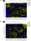
Overall, 102 antibodies were analyzed in 25 tumor samples, including five tumor types (a fresh frozen tissue; b FFPE specimens) with columns = tumor samples and rows = antibody signals. The color gradient from blue to yellow corresponds to low or high antibody-specific signals among the 25 tumor samples. COAD colorectal adenocarcinomas, HNSC head and neck squamous cell carcinomas, LUAD lung adenocarcinomas, LUSC lung squamous cell carcinomas, PAAD pancreatic adenocarcinomas.
Fig. 3
Fig. 3. Pairwise t-test performed for 102 antibodies for each pair of tumor types.
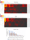
Heatmaps show p values (−log10 transformed) of the pairwise t-test (a fresh frozen and b FFPE tissue) after Benjamini–Hochberg (BH) correction. Columns represent the tested antibodies, and rows indicate pairs of tumor types. c Bar chart of the number of antibodies with a significant p value (p-BH < 0.05) in the pairwise t-test for each pair of tumors; comparison of fresh frozen samples, FFPE tissue, and overlap of both tissue types. COAD colorectal adenocarcinomas, HNSC head and neck squamous cell carcinomas, LUAD lung adenocarcinomas, LUSC lung squamous cell carcinomas, PAAD pancreatic adenocarcinomas.
Fig. 4
Fig. 4. Multiclass cancer classification by machine learning.
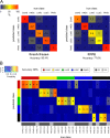
a Contingency matrices showing the classification accuracy of the SVM-based models obtained by repeated nested cross-validation with 25 tumor samples in fresh frozen and FFPE tissue. The numbers indicate how many of the five samples of each tumor type are classified on average in each class. The corresponding percentages are visualized by the color scheme (blue: low, red: high). b Classification results of the SVM algorithm in an independent validation cohort of 25 FFPE samples with five cases per tumor type. The predicted tumor type is marked with a cross; the color gradient indicates the confidence of the SVM classifier for each class (blue: low probability, red: high probability). Histological grades: G2 moderately and G3 poorly differentiated tumor samples. COAD colorectal adenocarcinomas, HNSC head and neck squamous cell carcinomas, LUAD lung adenocarcinomas, LUSC lung squamous cell carcinomas, PAAD pancreatic adenocarcinomas.

References

    1. Pavlidis N, Pentheroudakis G. Cancer of unknown primary site. Lancet. 2012;379:1428–35. - PubMed
    1. Economopoulou P, Mountzios G, Pavlidis N, Pentheroudakis G. Cancer of unknown primary origin in the genomic era: elucidating the dark box of cancer. Cancer Treat Rev. 2015;41:598–604. - PubMed
    1. Pentheroudakis G, Golfinopoulos V, Pavlidis N. Switching benchmarks in cancer of unknown primary: from autopsy to microarray. Eur J Cancer. 2007;43:2026–36. - PubMed
    1. Pereira TC, Share SM, Magalhães AV, Silverman JF. Can we tell the site of origin of metastatic squamous cell carcinoma? An immunohistochemical tissue microarray study of 194 cases. Appl Immunohistochem Mol Morphol. 2011;19:10–14. - PubMed
    1. Ramaswamy S, Tamayo P, Rifkin R, Mukherjee S, Yeang C-H, Angelo M, et al. Multiclass cancer diagnosis using tumor gene expression signatures. Proc Natl Acad Sci USA. 2001;98:15149–54. - PMC - PubMed

Publication types