. 2020 Jun 29;10(1):10556.
doi: 10.1038/s41598-020-67417-4.

Removal of an abluminal lining improves decellularization of human umbilical arteries

Affiliations

Removal of an abluminal lining improves decellularization of human umbilical arteries

Ho-Yi Tuan-Mu et al. Sci Rep. .

Abstract

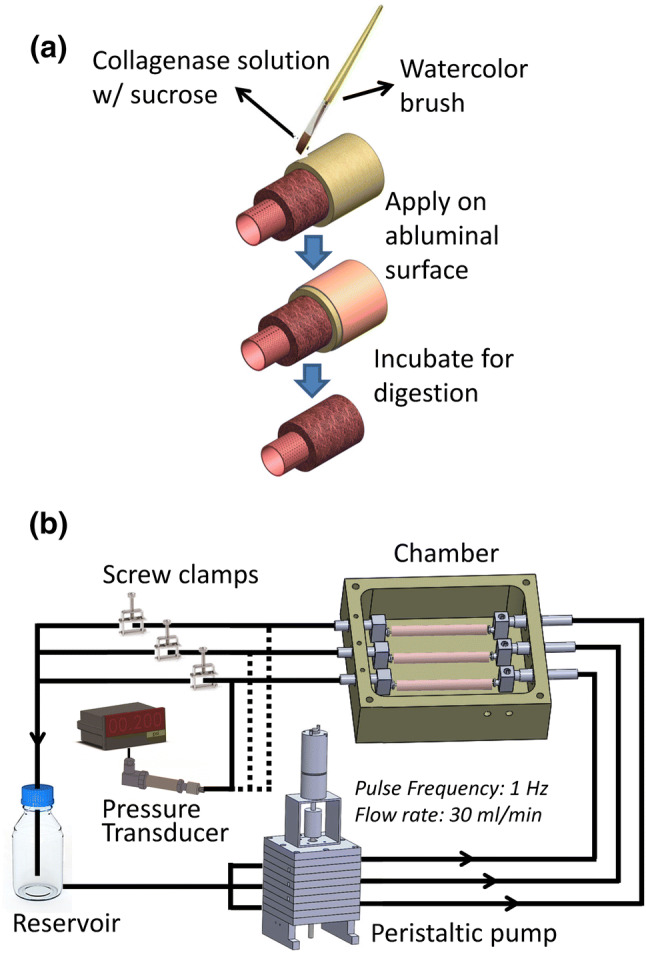
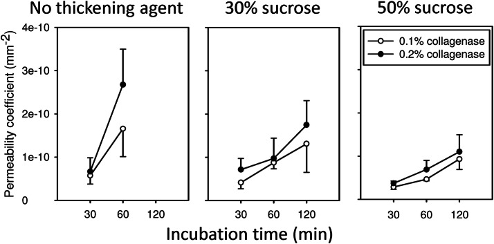
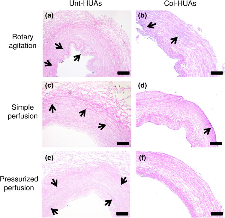
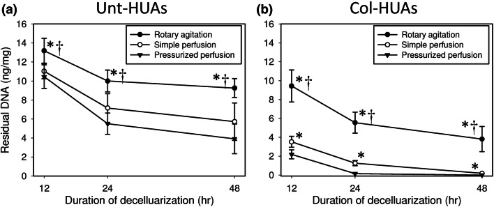
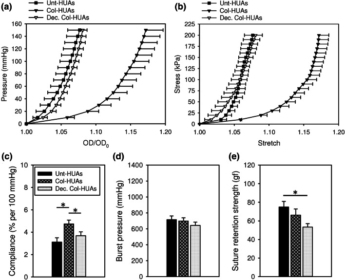
The decellularization of long segments of tubular tissues such as blood vessels may be improved by perfusing decellularization solution into their lumen. Particularly, transmural flow that may be introduced by the perfusion, if any, is beneficial to removing immunogenic cellular components in the vessel wall. When human umbilical arteries (HUAs) were perfused at a transmural pressure, however, very little transmural flow was observed. We hypothesized that a watertight lining at the abluminal surface of HUAs hampered the transmural flow and tested the hypothesis by subjecting the abluminal surface to enzyme digestion. Specifically, a highly viscous collagenase solution was applied onto the surface, thereby restricting the digestion to the surface. The localized digestion resulted in a water-permeable vessel without damaging the vessel wall. The presence of the abluminal lining and its successful removal were also supported by evidence from SEM, TEM, and mechanical testing. The collagenase-treated HUAs were decellularized with 1% sodium dodecyl sulfate (SDS) solution under either rotary agitation, simple perfusion, or pressurized perfusion. Regardless of decellularization conditions, the decellularization of HUAs was significantly enhanced after the abluminal lining removal. Particularly, complete removal of DNA was accomplished in 24 h by pressurized perfusion of the SDS solution. We conclude that the removal of the abluminal lining can improve the perfusion-assisted decellularization.


Conflict of interest statement

The authors declare no competing interests.

Figures

Figure 1
Figure 1
Schematics of the collagenase treatment procedures for removing the abluminal lining of HUAs (a) and of the perfusion system for decellularizing HUAs (b).
Figure 2
Figure 2
Time courses of permeability changes in the vessel wall of the HUAs treated with the collagenase solution containing no thickening agent (low viscosity), 30% sucrose (medium viscosity), or 50% sucrose (high viscosity). The HUAs treated with the collagenase solution of low viscosity for 120 min leaked tremendously when pressurized thus no permeability coefficient was calculated. Each error bar should be on both sides of the mean; only one side is shown for simplicity.
Figure 3
Figure 3
Representative SEM images of Col-HUA (a) and Unt-HUA (b,c) cross-sections. The arrows indicate the microstructure that was absent in Col-HUAs. Panel (c) shows higher magnification view of the box outlined in panel (b).
Figure 4
Figure 4
Representative TEM images of Col-HUA (a) and Unt-HUA (b,c) cross-sections near the abluminal surface. The arrows indicate the microstructure that was absent in Col-HUAs. Panel c shows higher magnification view of the box outlined in panel (b).
Figure 5
Figure 5
Average pressure-diameter curves (a) and circumferential stress-stretch curves (b) of Unt-HUAs and Col-HUAs. Data are presented as mean ± SD (n = 5).
Figure 6
Figure 6
Illustration of residual DNA in Unt-HUAs (a,c,e) and Col-HUAs (b,d,f) decellularized with 1% sucrose solution under rotary agitation, simple perfusion, or pressurized perfusion for 24 h with H&E stained histological sections. Arrows indicate nuclei debris.
Figure 7
Figure 7
Time course of residual DNA in Unt-HUAs (a) and Col-HUAs (b) decellularized with 1% SDS solution under rotary agitation, simple perfusion, or pressurized perfusion. Data are presented as mean ± SD (n = 5). *p < 0.025 vs. pressurized perfusion; p < 0.025 vs. simple perfusion.
Figure 8
Figure 8
Representative PSR stained (ac) and Alcian blue stained (eg) of Unt-HUAs (a,e), Col-HUAs (b,f), and decellularized Col-HUAs (c,g). Comparisons of relative collagen (d) and GAG (h) contents among the three groups. Data are presented as mean ± SD (n = 3). *p < 0.025. Scale bar 100 μm.
Figure 9
Figure 9
Average pressure-diameter curves (a) and circumferential stress-stretch curves (b) of Unt-HUA, Col-HUAs and decellularized Col-HUAs. (c) Comparisons of the compliance, burst pressure, and suture retention strength among Unt-HUAs, Col-HUAs, and decellularized Col-HUAs. Data are presented as mean ± SD (n = 5). *p < 0.05.

References

    1. Herrington W, Lacey B, Sherliker P, Armitage J, Lewington S. Epidemiology of atherosclerosis and the potential to reduce the global burden of atherothrombotic disease. Circ. Res. 2016;118:535–546. doi: 10.1161/Circresaha.115.307611. - DOI - PubMed
    1. Libby P, et al. Atherosclerosis. Nat. Rev. Dis. Primers. 2019;5:56. doi: 10.1038/s41572-019-0116-x. - DOI - PubMed
    1. Benjamin EJ, et al. Heart disease and stroke statistics-2017 update a report from the American Heart Association. Circulation. 2017;135:E146–E603. doi: 10.1161/Cir.0000000000000485. - DOI - PMC - PubMed
    1. Youngstrom DW, Barrett JG, Jose RR, Kaplan DL. Functional characterization of detergent-decellularized equine tendon extracellular matrix for tissue engineering applications. PLoS ONE. 2013;8:1. doi: 10.1371/journal.pone.0064151. - DOI - PMC - PubMed
    1. Grayson WL, et al. Engineering anatomically shaped human bone grafts. Proc. Natl. Acad. Sci. USA. 2010;107:3299–3304. doi: 10.1073/pnas.0905439106. - DOI - PMC - PubMed

Publication types