Review
. 2020 Aug;21(8):433-444.
doi: 10.1038/s41583-020-0325-z. Epub 2020 Jun 29.

Cellular senescence and Alzheimer disease: the egg and the chicken scenario

Affiliations
Review

Cellular senescence and Alzheimer disease: the egg and the chicken scenario

Sara Saez-Atienzar et al. Nat Rev Neurosci. 2020 Aug.

Erratum in

Abstract

Globally, 50 million people live with dementia, with Alzheimer disease (AD) being responsible for two-thirds of the total cases. As ageing is the main risk factor for dementia-related neurodegeneration, changes in the timing or nature of the cellular hallmarks of normal ageing might be key to understanding the events that convert normal ageing into neurodegeneration. Cellular senescence is a candidate mechanism that might be important for this conversion. Under persistent stress, as occurs in ageing, both postmitotic cells - including neurons - and proliferative cells - such as astrocytes and microglia, among others - can engender a state of chronic cellular senescence that is characterized by the secretion of pro-inflammatory molecules that promote the functional decline of tissues and organs. Ablation of senescent cells has been postulated as a promising therapeutic venue to target the ageing phenotype and, thus, prevent or mitigate ageing-related diseases. However, owing to a lack of evidence, it is not possible to label cellular senescence as a cause or a consequence of neurodegeneration. This Review examines cellular senescence in the context of ageing and AD, and discusses which of the processes - cellular senescence or AD - might come first.


Figures

Fig. 1 |
Fig. 1 |. The role of senescence in the context of ageing, Alzheimer disease and Alzheimer disease-related dementias.
Various processes may contribute to healthy ageing versus Alzheimer disease (AD)-related pathophysiology, with a focus on when senescence may play a role. In healthy individuals, there is a decline in the homeostasis capacity with ageing that contributes to the accumulation of different cellular stressors, such as oxidative stress, DNA damage and mitochondrial stress, among others. As a consequence, cells accumulate damage with ageing. Senescence may be activated as a homeostatic mechanism at first, but if senescent cells are not cleared, the accumulation of senescent cells becomes greater with time, triggering a chronic inflammatory response that contributes to a chronic low-grade inflammation (inflammaging) and that participates in tissue decline. Inflammation induces synapse damage and contributes to cognitive decline. Individuals with AD, including individuals carrying genetic mutations associated with the disease, are exposed to additional internal stressors associated with neurodegeneration that will trigger proteinopathy. Recent data show that toxic protein aggregation, such as tau aggregation and amyloid plaque deposition,, acts as a pro-senescence stimulus and induces cellular senescence in different brain cell types, leading to local inflammation in the tissues targeted by the disease. This inflammation, in combination with the toxic protein aggregation, contributes to a greater accumulation of stressors within the cells. The resulting cellular damage, in combination with the local senescence, boosts the ageing phenotype and contributes to chronic senescence. There is a third scenario, also associated with the role of senescence in AD and AD-related dementias (ADRDs), in which other proteinopathy-independent mechanisms might trigger senescence activation. This proposal is based on previous work showing that the formation of senescent cells precedes tau aggregation in the MAPTP301SPS19 mouse model of tauopathy (REF.) but it has not been widely explored yet in AD. In this proposal, factors such as excessive myelin fragmentation or a higher burden of genetic risk variants for cellular senescence could induce senescence in the absence of proteinopathy. The accumulation of tissue-specific senescence could generate a harmful environment for cells, making them more susceptible to toxic protein aggregation, and induce proteinopathy, both contributing to chronic senescence and boosting neural loss and cognitive decline. ROS, reactive oxygen species.
Fig. 2 |
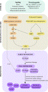
Fig. 2 |. Cellular mechanisms and phenotypic features of senescent cells in ageing and neurodegeneration.
Cellular senescence is a complex process that is triggered by a myriad of pro-senescent stimuli related with ageing, such as reactive oxygen species (ROS), telomere attrition, lysosomal dysfunction and mitochondrial alterations, among others. Recently, Alzheimer disease (AD)-related proteinopathy events have also been related to senescence induction, such as amyloid deposition and tau aggregation. TAR DNA-binding protein 43 (TDP43) and α-synuclein might also be potential candidates to induce senescence, but there are still no publications that support this claim. DNA damage is one of the main signals that induces cellular senescence activation. DNA damage actives the DNA damage response (DDR) machinery, responsible for p53–p21Cip1 axis activation, which in turn blocks cyclin-dependent kinase 2 (CDK2) activity, resulting in retinoblastoma (RB) hypophosphorylation and cell cycle exit. INK4–ARF locus de-repression is another senescence activation mechanism. The INK4–ARF locus is repressed in normal cells by polycomb proteins and epigenetic factors, but when it is activated, it promotes the expression of ARF (murine p19ARF and human p14ARF), which is known to prevent p53 degradation, and the expression of p16INK4a, which consequently inhibits CDK4 and CDK6 and leads to RB hypophosphorylation and long-lasting arrest of the cell cycle. DNA damage and INK4–ARF locus de-repression converge in RB hypophosphorylation, and it is thought that the two paths act together to induce and maintain senescence in ageing and neurodegeneration. As a consequence of the cell cycle arrest, cells enter into an early senescence stage that evolves to a full senescence, characterized by the senescence-associated secretory phenotype (SASP). The DDR regulates the induction of SASP through the activation of the transcription factor NF-κB. In addition, epigenetic regulation acts at the promoters of the genes encoding interleukin 6 (IL-6) and IL-8, both of which are constituents of the SASP. If fully senescent cells are not cleared, they evolve to a state of late senescence (also known as chronic senescence). The accumulation of senescent cells in this stage occurs in ageing and neurodegeneration and leads to chronic inflammation, which contributes to organ and tissue decline. Three main factors contribute to late senescence transition: first, sustained and chronic damage; second, a reduction in the capacity of the immune system to remove senescent cells in aged individuals; and, third, the capacity of senescent cells to induce senescence in their surroundings. ATM, ataxia telangiectasia mutated; ATR, ataxia telangiectasia and Rad3-related protein; SA-β-gal, senescence-associated β-galactosidase.
Fig. 3 |
Fig. 3 |. Alzheimer disease pathology in the context of myelin fragmentation and senescence.
Alzheimer disease (AD) is characterized by the accumulation of intracellular abnormal tau and extracellular amyloid plaques. Myelin loss is an early event in the pathobiology of AD and is enhanced by the presence of extracellular amyloid plaques. Microglial cells act as macrophages and they phagocytose the myelin debris and then degrade it through the autophagy–lysosomal pathway. When the accumulation of myelin debris is greater than the microglial lysosomal degradative capacity, microglial cells become senescent and release proinflammatory factors (collectively referred to as the senescence-associated secretory phenotype (SASP)). We propose that the activation of senescence in microglial cells has two negative outcomes that may contribute directly to the pathology observed in AD: inflammation activation, which contributes to the impairment of other cell types, such as oligodendrocytes and oligodendrocyte progenitor cells (OPCs); and loss of microglial capacity, which contributes to a greater accumulation of amyloid plaques and myelin debris. In addition, the accumulation of myelin debris could impair the remyelination process by affecting oligodendrocyte recruitment to the axons and by suppressing microglial-derived factors that are required for OPC differentiation into oligodendrocytes.

References

    1. Wortmann M World Alzheimer report 2014: Dementia and risk reduction. Alzheimer’s & Dementia vol. 11 P837 (2015).
    1. Winblad B et al. Defeating Alzheimer’s disease and other dementias: a priority for European science and society. Lancet Neurol. 15, 455–532 (2016). - PubMed
    1. Ritchie K & Lovestone S The dementias. The Lancet vol. 360 1759–1766 (2002). - PubMed
    1. Corriveau RA et al. Alzheimer’s Disease-Related Dementias Summit 2016: National research priorities. Neurology 89, 2381–2391 (2017). - PMC - PubMed
    1. Hou Y et al. Ageing as a risk factor for neurodegenerative disease. Nat. Rev. Neurol. 15, 565–581 (2019). - PubMed

Publication types