. 2020 Jun 29;10(1):10518.
doi: 10.1038/s41598-020-67136-w.

AAV8 Ins1-Cre can produce efficient β-cell recombination but requires consideration of off-target effects

Affiliations

AAV8 Ins1-Cre can produce efficient β-cell recombination but requires consideration of off-target effects

Adam Ramzy et al. Sci Rep. .

Abstract

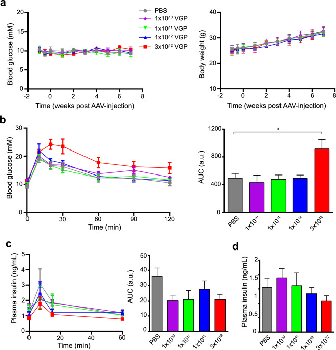
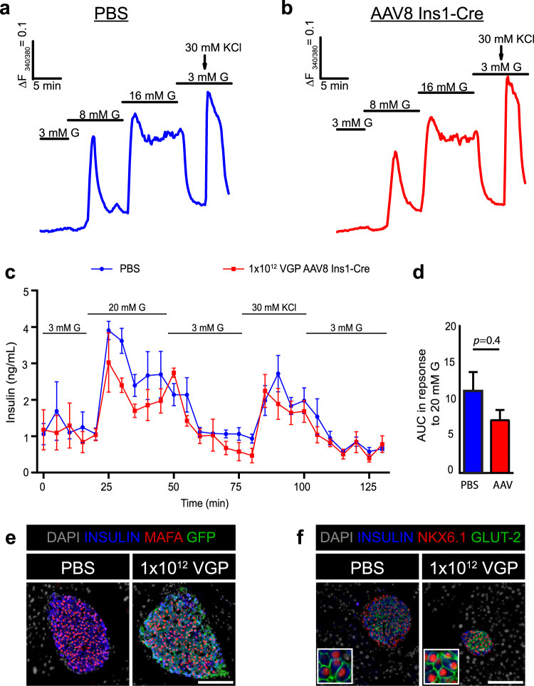
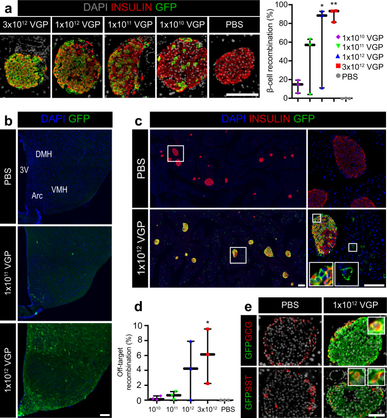
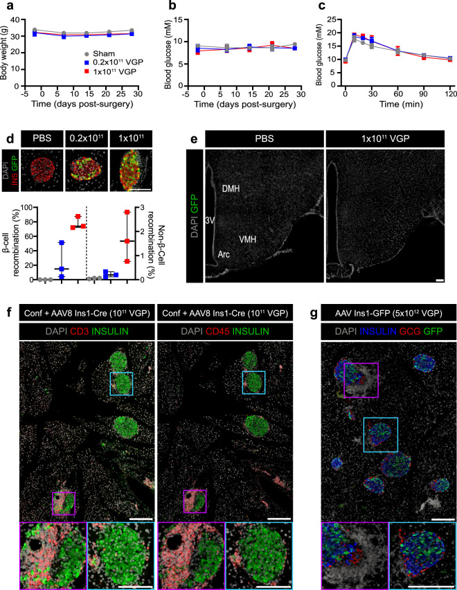
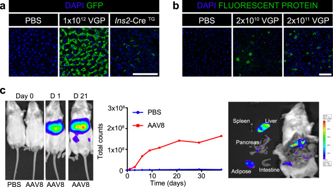
In vivo genetic manipulation is used to study the impact of gene deletion or re-expression on β-cell function and organism physiology. Cre-LoxP is a system wherein LoxP sites flanking a gene are recognized by Cre recombinase. Cre transgenic mice are the most prevalent technology used to deliver Cre but many models have caveats of off-target recombination, impaired β-cell function, and high cost of animal production. Inducible estrogen receptor conjugated Cre models face leaky recombination and confounding effects of tamoxifen. As an alternative, we characterize an adeno associated virus (AAV) with a rat insulin 1 promoter driving Cre recombinase (AAV8 Ins1-Cre) that is economical and rapid to implement, and has limited caveats. Intraperitoneal AAV8 Ins1-Cre produced efficient β-cell recombination, alongside some hepatic, exocrine pancreas, α-cell, δ-cell, and hypothalamic recombination. Delivery of lower doses via the pancreatic duct retained good rates of β-cell recombination and limited rates of off-target recombination. Unlike inducible Cre in transgenic mice, AAV8 Ins1-Cre required no tamoxifen and premature recombination was avoided. We demonstrate the utility of this technology by inducing hyperglycemia in inducible insulin knockout mice (Ins1-/-;Ins2f/f). AAV-mediated expression of Cre in β-cells provides an effective alternative to transgenic approaches for inducible knockout studies.


Conflict of interest statement

The authors declare no competing interests.

Figures

Figure 1
Figure 1
Intraperitoneal administration of AAV8 Ins1-Cre up to a single dose of 1 × 10¹² VGP does not significantly alter glucose metabolism. (a) 4-hour fasting blood glucose and body weight of adult mTmG reporter mice injected with either vehicle (PBS) or different doses of AAV8 Ins1-Cre. (b) Oral glucose tolerance test (6 hour fast, 2 g glucose/kg body weight) 7 weeks post-virus injection. Area under the curve (AUC) relative to baseline is presented to the right in arbitrary units (a.u.). (c) Plasma insulin levels during oral glucose tolerance test and (d) after 6 hours fasting 8 weeks post-virus injection. Data are expressed as mean ± SEM and were analyzed using one- or two-way repeated measures ANOVA. n = 4–5 mice per group. (*p < 0.05).
Figure 2
Figure 2
Intraperitoneal administration of AAV8 Ins1-Cre (1 × 10¹² VGP) does not significantly alter β-cell maturity or function. Adult C57BL/6 J mice were given either PBS or 1 × 10¹² VGP of AAV8 Ins1-Cre by single intraperitoneal injection, and islets were isolated three to four weeks post-injection. Representative [Ca2+]i recordings of an islet from a PBS treated mouse (a) and an AAV8 Ins1-Cre injected mouse (b), in response to different glucose (G) concentrations and potassium chloride (KCl). Graphs are representative of 31–47 islets from 3 mice per group. (c) Insulin secretion in response to G and KCl in islets from PBS and AAV-injected mice, and (d) area under the curve (AUC) in response to high glucose (20 mM G) from islets perifused in (c). Batches of 80 islets from 3 mice per group were employed. Mann-Whitney test was performed. Pancreas from adult mTmG mice injected with PBS or AAV8 Ins1-Cre was immunostained for mature β-cell factors MAFA (e), NKX6.1, and GLUT-2 (f). Representative images of n = 3 shown. Scale bars are 100 μm.
Figure 3
Figure 3
Intraperitoneal AAV8 Ins1-Cre produces dose-dependent β-cell recombination alongside hypothalamic and acinar tissue recombination. Pancreas from mTmG mice administered PBS or varying doses of AAV8 Ins1-Cre was collected 10 weeks post-AAV and immunostained for insulin and GFP (a), and recombination rate in β-cells was quantified (n = 3). Groups were compared to PBS control by Kruskal-Wallis test with Dunn’s post hoc test. (b) Brains from these mice were immunostained for GFP with nuclear DAPI counterstain. A labeled coronal section through the mid-hypothalamus is shown (3 V: third ventricle, ARC: arcuate nucleus, VMH: ventromedial nucleus of the hypothalamus, and DMH: dorsomedial nucleus of the hypothalamus). Representative image of n = 3. (c,d) Significant exocrine and insulin negative islet cell recombination in AAV8 Ins1-Cre injected mice. Representative images of n = 3 shown. Quantification of the proportion of insulin negative cell recombination was compared to PBS control by one-way ANOVA with Tukey’s post-hoc test. (e) Pancreas was immunostained for GFP and the islet hormones glucagon (GCG) or somatostatin (SST). Representative images of n = 3 shown. Scale bars in all panels are 100 μm. Insets are enlarged 4×. (*p < 0.05, **p < 0.01).
Figure 4
Figure 4
AAV8 Ins1-Cre intraductal administration does not significantly alter glucose tolerance but induction of foreign fluorescent protein expression causes insulitis. Four hour fasting body weight (a) and blood glucose (b) of adult confetti reporter mice that received AAV8 Ins1-Cre by intraductal delivery. (c) Oral glucose tolerance test (2 g glucose/kg body weight) 4 weeks post virus administration. Data are expressed as mean ± SEM and were analyzed using two-way repeated measures ANOVA. n = 7–8 mice in each group (ac). Pancreas was collected seven weeks post-AAV and immunostained for insulin (INS) and GFP, and proportion of INS+ or INS- cells that were GFP + were quantified (d). Representative images of n = 3 shown and individual animal data points shown. Groups were compared to PBS control by Kruskal-Wallis test with Dunn’s post hoc test. (e) Brains were immunostained for GFP and a coronal section through the mid-hypothalamus is shown (3 V: third ventricle, ARC: arcuate nucleus, VMH: ventromedial nucleus of the hypothalamus, and DMH: dorsomedial nucleus of the hypothalamus). Representative image of n = 1 of control or n = 3 of 1 × 10¹¹ VGP injected mice shown. There was no detectable GFP in 0.2 × 10¹¹ VGP dosed mice (data not shown). (f) Adult confetti mice injected with AAV8 Ins1-Cre had insulitis with CD3 + and CD45 + cells surrounding islets. Representative images of n = 3 shown. (g) Adult C57BL/6 J mice were injected with 5 × 10¹² VGP AAV8 Ins1-GFP IP and developed insulitis by 4 months post-AAV. Representative images of n = 2. Scale bars are 100 μm.
Figure 5
Figure 5
Both IP and ID administration of AAV8 Ins1-Cre can cause recombination in the liver but the extent may be dependent on delivery method and mouse strain. Liver from adult mTmG mice given IP AAV8 Ins1-Cre (a) or confetti mice given ID AAV8 Ins1-Cre (b) was immunostained for fluorescent proteins to identify hepatic recombination events. Representative images of n = 2–3 shown. Scale bars are 100 μm. (c) Adult Rosa26-LSL-luciferase mice were administered 1 × 10¹² VGP AAV8 Ins1-Cre IP (n = 3) or PBS (n = 1) and luciferase activity was assessed following luciferin injection for 5 weeks including after rapid dissection at study termination.
Figure 6
Figure 6
The AAV8 Ins1-Cre can be a useful tool for directing β-cell recombination when off-target effects are deemed minimally important. Four hour fasting body weight (a) and blood glucose (b) of adult Ins1−/−;Ins2f/f mice that received AAV8 Ins1-Cre by IP delivery (indicated by black arrow). IPGTTs were performed on day 10 (c) and day 28 (d) relative to AAV injection. (e) Blood collected during the IPGTT on day 28 was assayed for circulating insulin. (f) Fasting insulin levels throughout the study were assessed by ELISA. Data shown as mean ± SEM (af) with individual traces (bf). Pancreata collected six weeks post-AAV were immunostained for insulin and Cre (g). Insets show 4x enlargement of representative cells with bright insulin and Cre immunoreactivity, diminished insulin and Cre immunoreactivity, and cells in the core of the islet with neither insulin nor Cre immunoreactivity. (h) Pancreas was immunostained for insulin and IAPP. The intensity of insulin immunoreactivity in IAPP + cells was quantified, and individual cells for each animal are presented as violin plots with percent of cells below a designated cut-off (black line) shown as mean ± 95% confidence interval. IAPP immunoreactivity was used to calculate the β-cell area as a percent of total pancreas area and average islet β-cell area. (i) Pancreas was immunostained for insulin and CD3. Data analysed by repeated measures two-way ANOVA (af) or Mann-Whitney test (h). n = 4–5, scale bars are 100 μm, and insets are enlarged 4×. (**p < 0.01, ***p < 0.001).

References

    1. Gu H, Marth JD, Orban PC, Mossmann H, Rajewsky K. Deletion of a DNA polymerase beta gene segment in T cells using cell type-specific gene targeting. Science. 1994;265:103–106. doi: 10.1126/science.8016642. - DOI - PubMed
    1. Magnuson MA, Osipovich AB. Pancreas-specific Cre driver lines and considerations for their prudent use. Cell Metab. 2013;18:9–20. doi: 10.1016/j.cmet.2013.06.011. - DOI - PMC - PubMed
    1. Postic C, et al. Dual roles for glucokinase in glucose homeostasis as determined by liver and pancreatic beta cell-specific gene knock-outs using Cre recombinase. J Biol Chem. 1999;274:305–315. doi: 10.1074/jbc.274.1.305. - DOI - PubMed
    1. Lee JY, et al. RIP-Cre revisited, evidence for impairments of pancreatic beta-cell function. J Biol Chem. 2006;281:2649–2653. doi: 10.1074/jbc.M512373200. - DOI - PubMed
    1. Teitelman G, Kedees M. Mouse insulin cells expressing an inducible RIPCre transgene are functionally impaired. J Biol Chem. 2015;290:3647–3653. doi: 10.1074/jbc.M114.615484. - DOI - PMC - PubMed

Publication types

Grants and funding