. 2020 Aug;21(8):857-867.
doi: 10.1038/s41590-020-0705-6. Epub 2020 Jun 29.

Ancient familial Mediterranean fever mutations in human pyrin and resistance to Yersinia pestis

Affiliations

Ancient familial Mediterranean fever mutations in human pyrin and resistance to Yersinia pestis

Yong Hwan Park et al. Nat Immunol. 2020 Aug.

Abstract

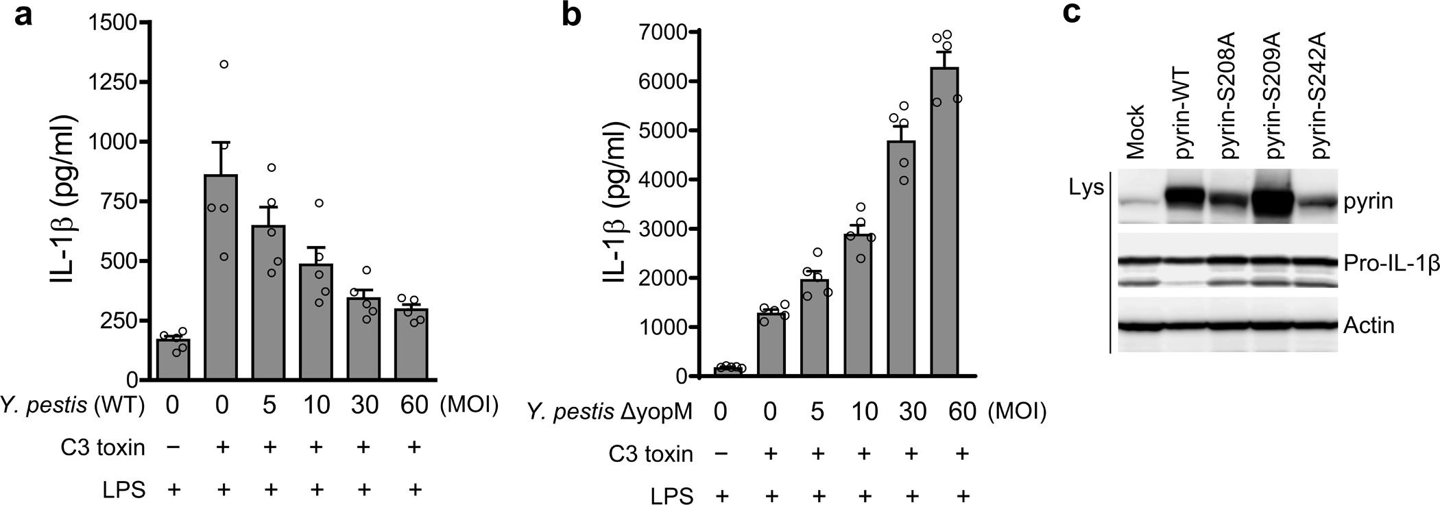
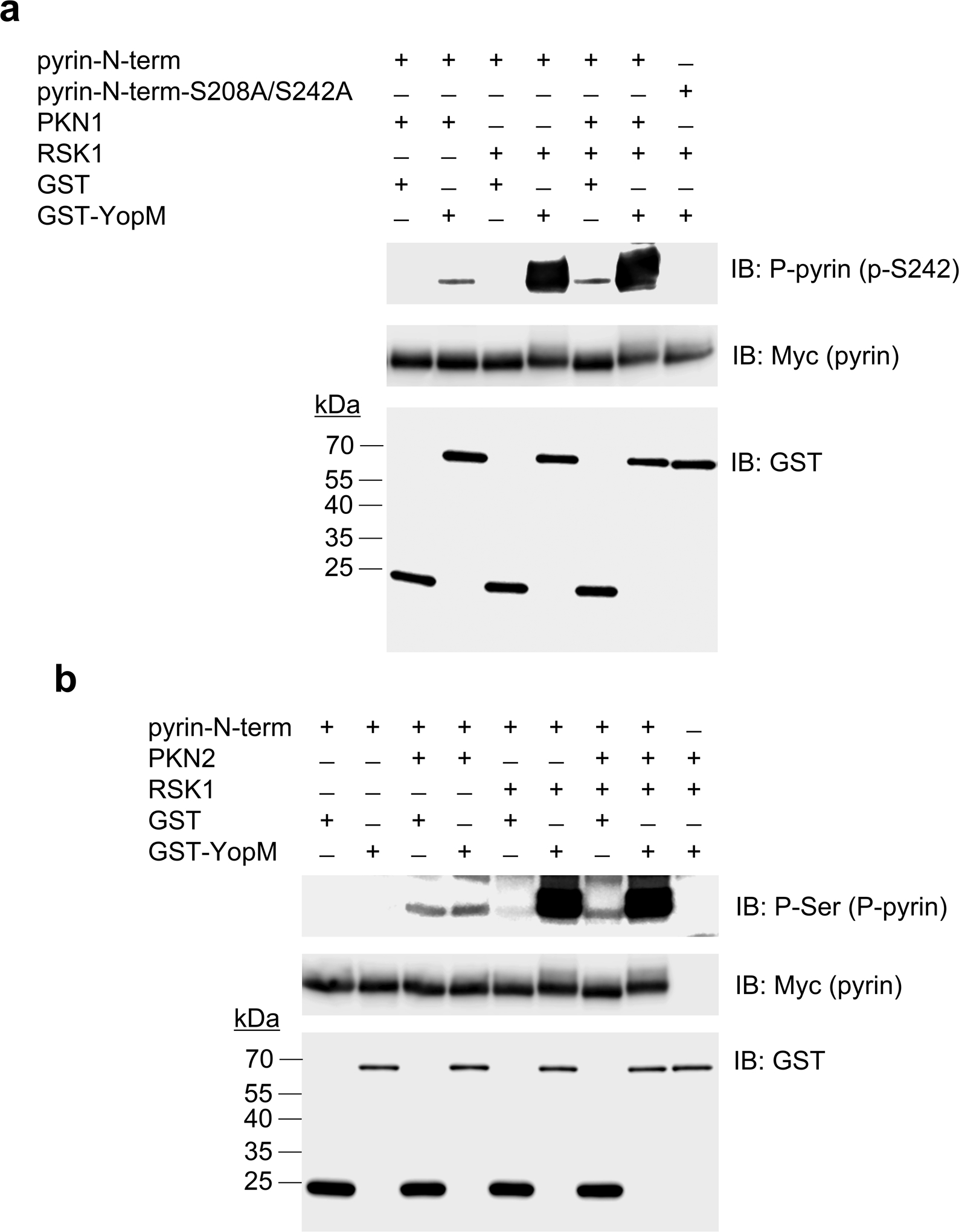
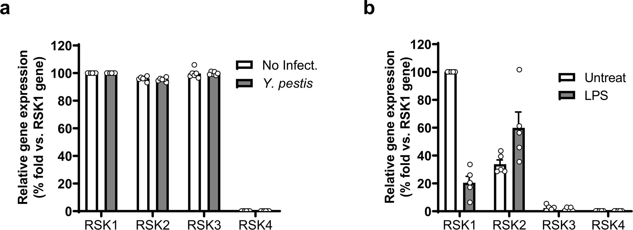
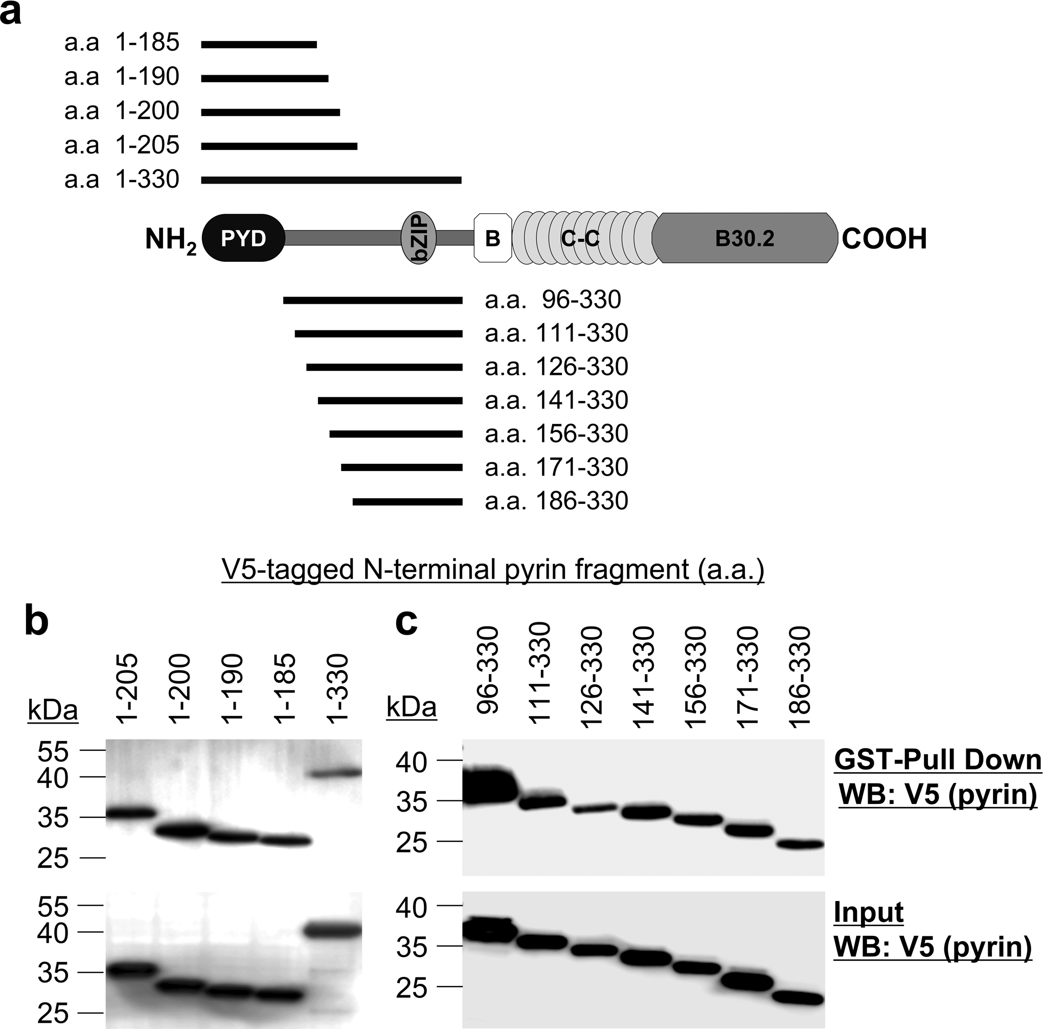
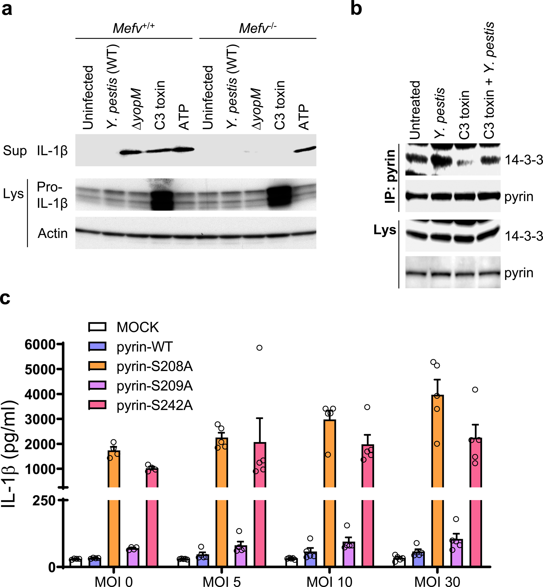
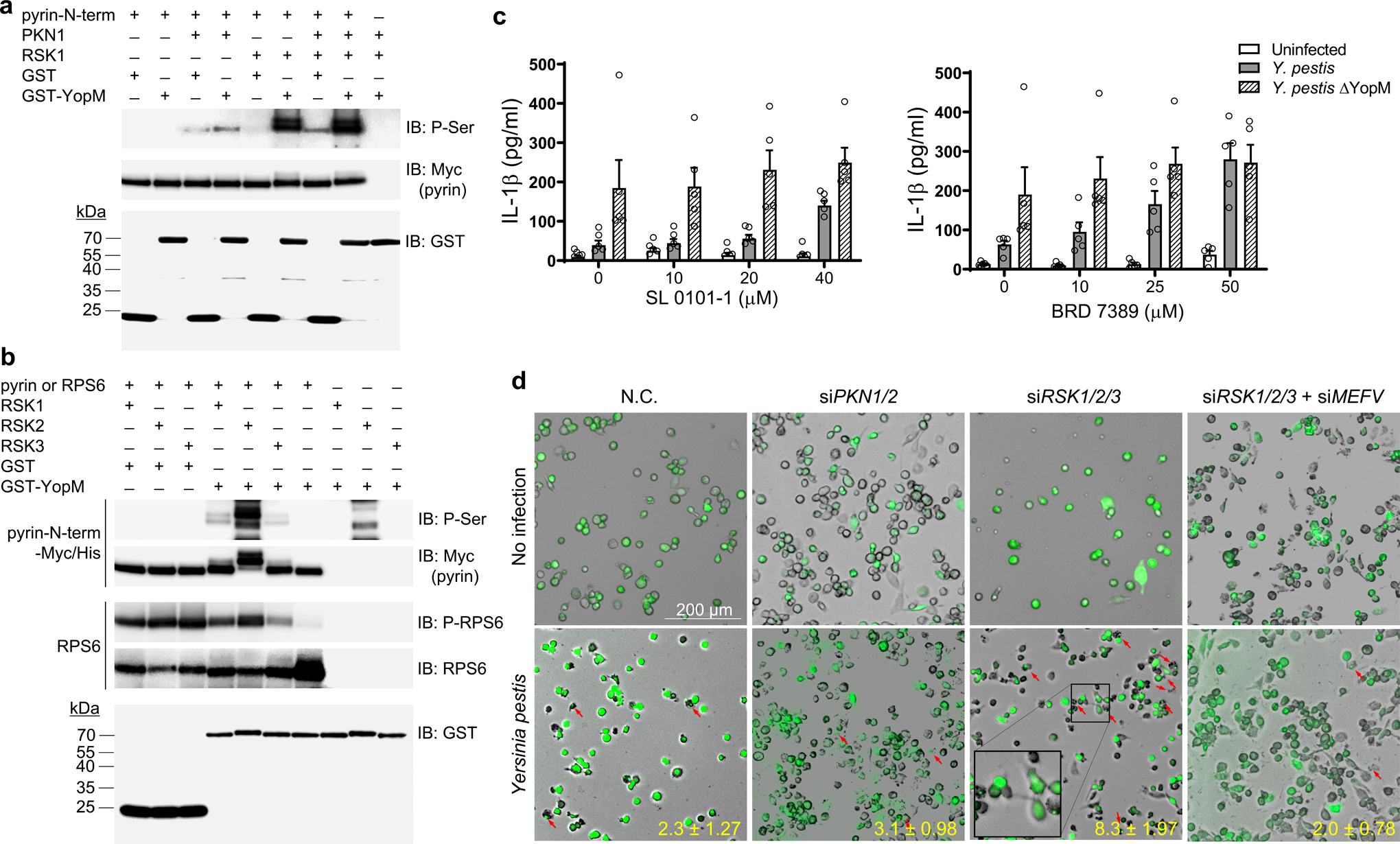
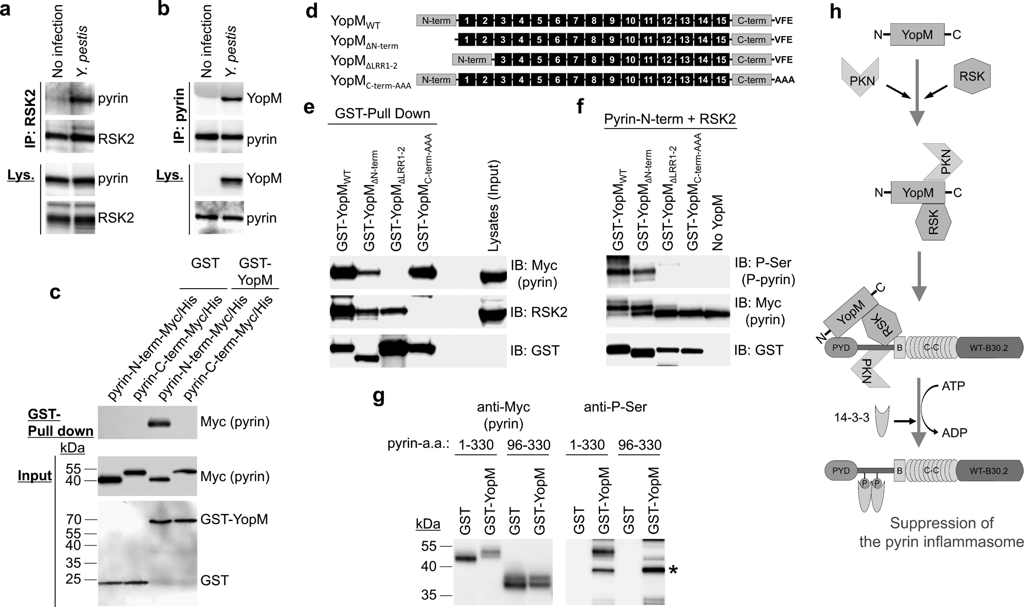
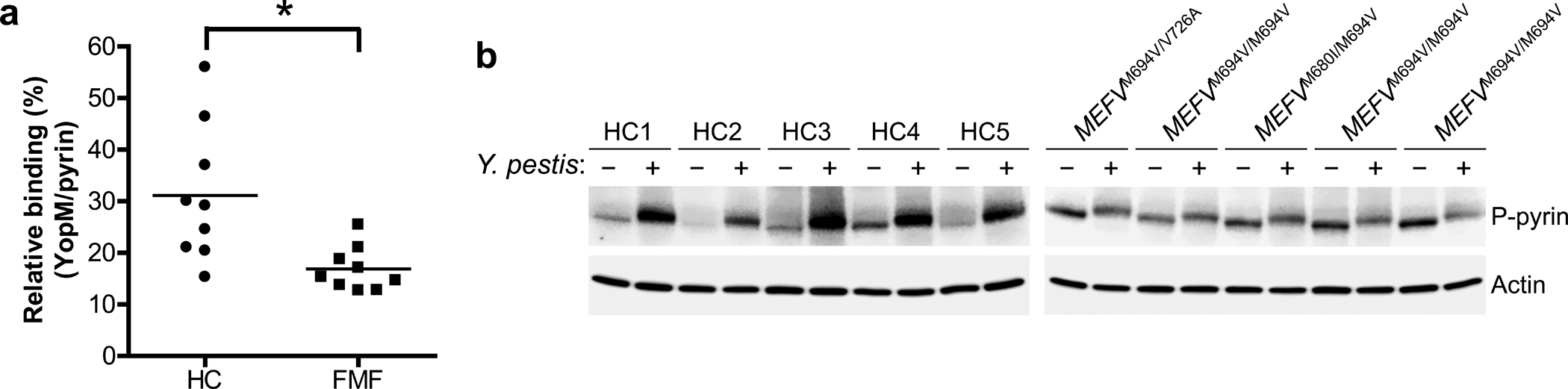
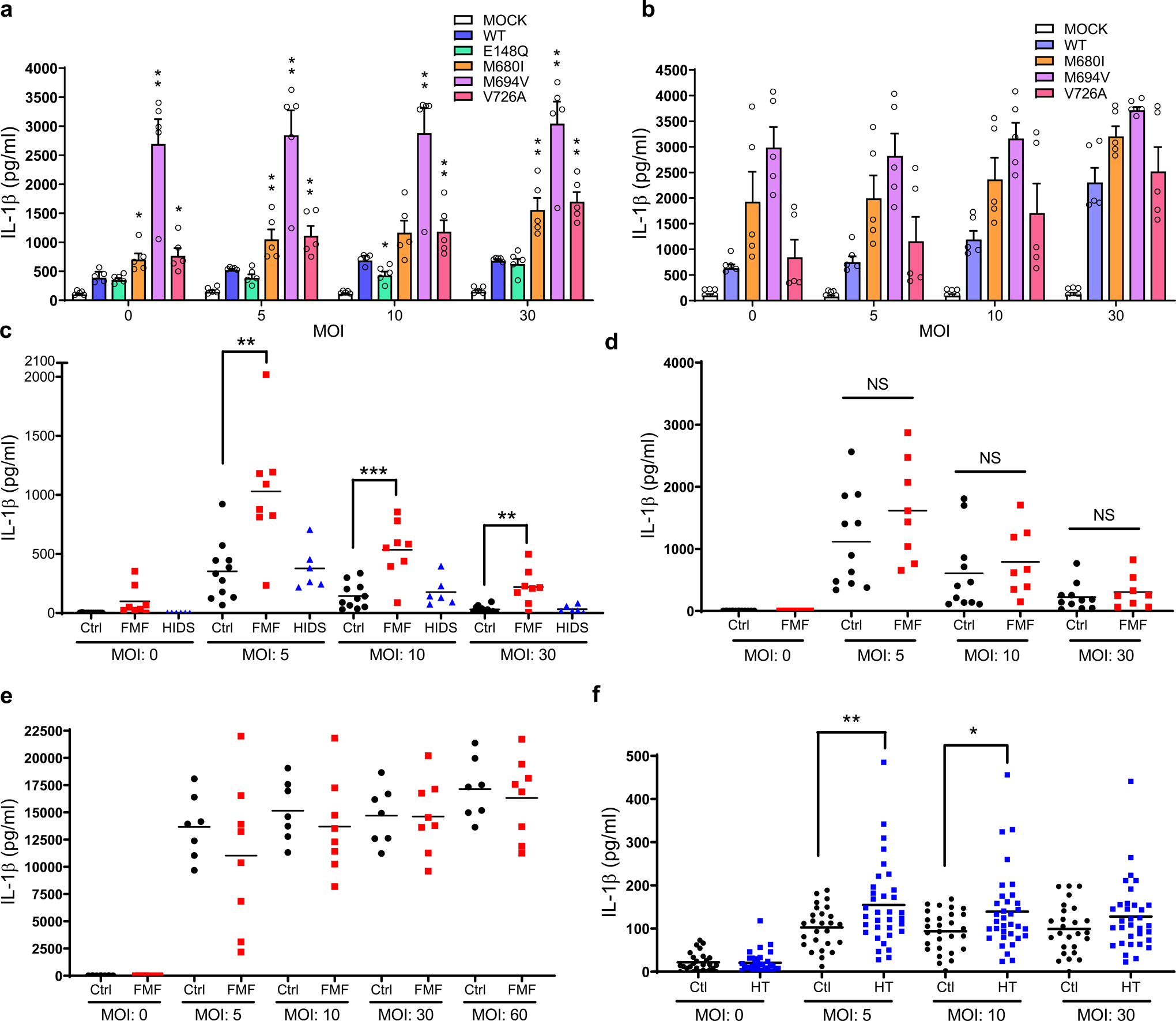
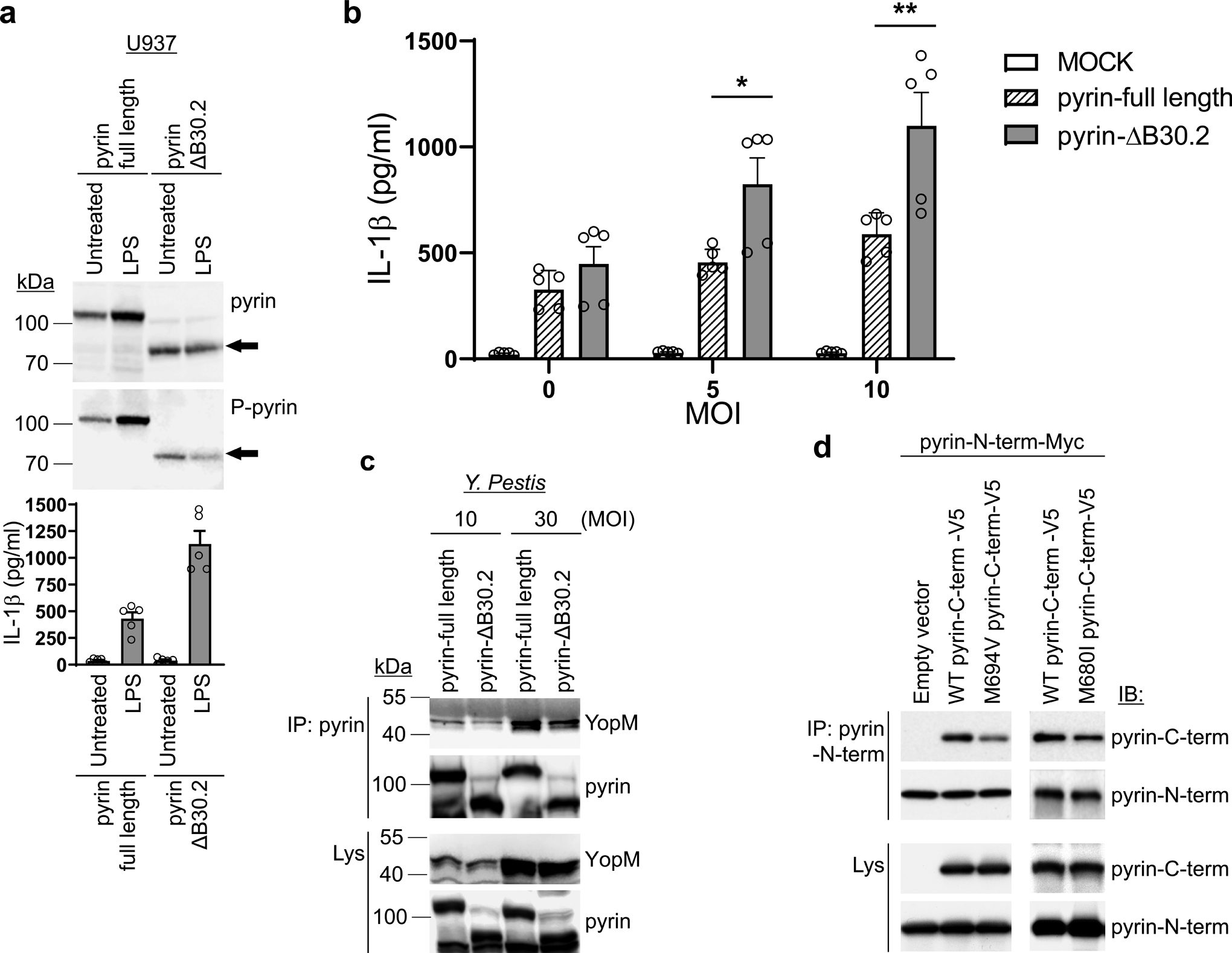
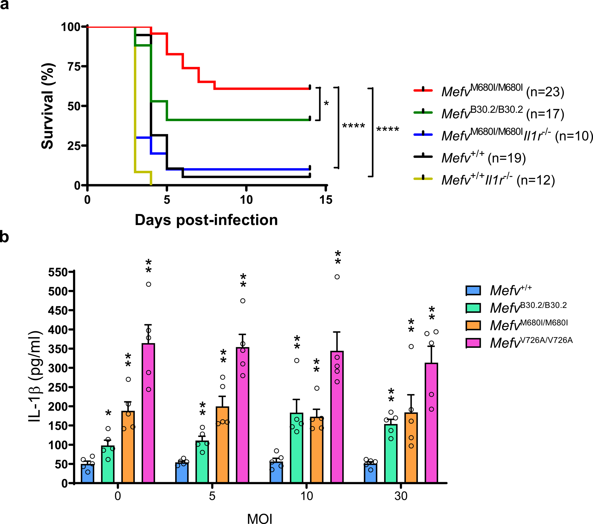
Familial Mediterranean fever (FMF) is an autoinflammatory disease caused by homozygous or compound heterozygous gain-of-function mutations in MEFV, which encodes pyrin, an inflammasome protein. Heterozygous carrier frequencies for multiple MEFV mutations are high in several Mediterranean populations, suggesting that they confer selective advantage. Among 2,313 Turkish people, we found extended haplotype homozygosity flanking FMF-associated mutations, indicating evolutionarily recent positive selection of FMF-associated mutations. Two pathogenic pyrin variants independently arose >1,800 years ago. Mutant pyrin interacts less avidly with Yersinia pestis virulence factor YopM than with wild-type human pyrin, thereby attenuating YopM-induced interleukin (IL)-1β suppression. Relative to healthy controls, leukocytes from patients with FMF harboring homozygous or compound heterozygous mutations and from asymptomatic heterozygous carriers released heightened IL-1β specifically in response to Y. pestis. Y. pestis-infected MefvM680I/M680I FMF knock-in mice exhibited IL-1-dependent increased survival relative to wild-type knock-in mice. Thus, FMF mutations that were positively selected in Mediterranean populations confer heightened resistance to Y. pestis.


Conflict of interest statement

COMPETING INTERESTS

The authors declare no competing interests

Figures

Extended Data Fig. 1
Extended Data Fig. 1. Haplotypes from 2,313 Turkish individuals provide evidence of evolutionarily recent positive selection and estimates of selection intensity and duration of selection on FMF-associated pyrin mutations.
a-c, Histograms of unstandardized nSL statistics of GWAS variants with similar frequency and local recombination rate as each FMF mutation. a, 1,402 markers similar to MEFV_p.V726A; b, 2,380 markers similar to MEFV_p.M694V; and c, 5,442 markers similar to MEFV_p.E148Q. The most extreme negative nSL values have the strongest evidence of recent positive selection. d-f, Log-likelihood plots and selection co-efficient estimates with 95% confidence intervals and mutation age estimates with 95% confidence intervals of three MEFV mutations, determined with a hidden Markov model method from the multi-locus haplotype structure of 4,626 Turkish chromosomes. d, MEFV_p.V726A; e, MEFV_p.M694V; and f, MEFV_p.E148Q. g-i, Histograms of iHH values for the ancestral alleles of markers with similar allele frequency and local recombination rate as each MEFV mutation, demonstrating that the ancestral alleles have not been under positive selection, thereby providing evidence that the mutations have not been under balancing selection. g, 1,570 markers similar to MEFV_ p.V726, h, 2,677 markers similar to MEFV_p.M694, and i, 6,166 markers similar to MEFV_p.E148.
Extended Data Fig. 2
Extended Data Fig. 2. Geographically diverse FMF mutation carriers exhibit extended conserved haplotypes.
a, MEFV_p.M694V conserved haplotypes (yellow). The length of the MEFV_p.M694V haplotype shared by all the diverse carrier chromosomes = 48 kb and shared by greater than 50% of the diverse carrier chromosomes = 307 kb. b, MEFV_p.V726A conserved haplotypes (pink). The length of the MEFV_p.V726A haplotype shared by all the diverse carrier chromosomes = 77 kb and shared by greater than 50% of the diverse carrier chromosomes = 262 kb. The fully conserved shared haplotypes in the diverse mutation carriers are identical to the respective haplotypes in the Turkish population.
Extended Data Fig. 3
Extended Data Fig. 3. Y. pestis YopM suppresses the pyrin inflammasome.
a,b, IL-1β measurements of culture supernatants of Mefv+/+ BMDMs primed with LPS, treated with or without C3 toxin, and infected with indicated MOI of WT (a) or ΔyopM (b) Y. pestis strains. Results are presented as mean ± s.e.m., for n=5 independent biological replicates. c, Immunoblot analysis of pyrin and pro-IL-1β in lysates of retroviral transduced U937 cells, expressing WT or indicated Ser to Ala mutant pyrin proteins. Data are representative of three independent experiments with similar results.
Extended Data Fig. 4
Extended Data Fig. 4. RSK-YopM mediated pyrin phosphorylation is independent of PKN.
In vitro kinase assay of purified Myc/His-tagged N-terminal human pyrin (amino acids 1–330) incubated with recombinant PKN1 and/or RSK1 (a) or PKN2 and/or RSK1 (b) in the presence of purified GST or GST-YopM, and analyzed by immunoblot with antibody specific for S242 phosphorylated pyrin (a) or phosphorylated serine (b). Data are representative of three independent experiments with similar results.
Extended Data Fig. 5
Extended Data Fig. 5. Human and murine RSK isoforms exhibit a restricted distribution of gene expression in leukocytes.
5 a,b, Relative gene expression profile of RSK isoforms in human PBMCs with or without Y. pestis infection (a) and mouse BMDMs with or without LPS priming (b), and assayed by RT-QPCR. Results are presented as mean ± s.e.m., for n=5 independent biological replicates.
Extended Data Fig. 6
Extended Data Fig. 6. Knockdown of RSK1, RSK2 and RSK3 in THP-1 cells induces ASC oligomerization.
a, Immunoblot analysis of lysates of THP-1 cells transiently transfected with negative control siRNA with no substantial sequence similarity to human gene sequences (N.C.) or a mixture of siRNAs targeting RSK1, RSK2 and RSK3. b, ASC oligomerization analysis by immunoblot from THP-1 cells transfected with N.C. or a mixture of siRNAs targeting RSK1, RSK2 and RSK3, and infected with Y. pestis (MOI 30). Cell lysates and the disuccinimidyl suberate (DSS)-treated pellets were analyzed by immunoblot with anti-ASC antibody. Data are representative of three independent experiments with similar results.
Extended Data Fig. 7
Extended Data Fig. 7. YopM binds to multiple regions of N-terminal pyrin.
a, Schematic structure of human pyrin with various deletion fragments of N-terminal human pyrin. b,c, GST-pulldown assay of V5-tagged various N-terminal human pyrin fragments with purified GST-YopM. Data are representative of three independent experiments with similar results.
Figure 1.
Figure 1.. Turkish population haplotypes provide evidence of evolutionarily recent positive selection of FMF-associated pyrin mutations.
a, Extended linkage disequilibrium (LD) surrounding MEFV_p.V726A and MEFV_p.M694V, but not MEFV_p.E148Q, in 2,313 Turkish individuals. (LD depicted by the four-gamete rule with black boxes indicating pairs of markers for which the 4th (recombinant) gamete frequency is less than 0.003, Haploview). b, Extended haplotype homozygosity (EHH) of haplotypes bearing FMF mutations compared with haplotypes with the ancestral alleles determined in 4,626 Turkish haplotypes. c, Histograms of unstandardized iHS statistics of GWAS markers with similar frequency and local recombination rate as each FMF mutation. Top panel, 1,570 markers similar to MEFV_p.V726A; middle panel, 2,677 markers similar to MEFV_p.M694V; bottom panel, 6,166 markers similar to MEFV_p.E148Q. The most extreme negative iHS values have the strongest evidence of recent positive selection. d, Representative trajectories from forward-time simulation of episodic selection. The depicted trajectories reflect a single run of the simulated evolutionary process. The plague pandemics are depicted by the hatched rectangles. The red curve depicts a representative trajectory for MEFV_p.M694V from one initial allelic copy 197 generations ago to an allele frequency of 2.1×10⁻³ at the beginning of the first plague pandemic and to a present-day allele frequency of 0.0313. The blue curve depicts a representative trajectory for MEFV_p.V726A from one initial allelic copy 163 generations ago to 1.6×10⁻³ at the beginning of the first plague pandemic and to a present-day allele frequency of 0.0197.
Figure 2
Figure 2. The pyrin inflammasome is suppressed by Y. pestis YopM through phosphorylation and subsequent 14-3-3 binding.
a, Immunoblot analysis of IL-1β in culture supernatants (Sup) and lysates (Lys) of WT (Mefv+/+) or Mefv−/− mouse BMDMs primed with LPS and treated with either C3 toxin or ATP, or infected with WT or ΔyopM Y. pestis strains at MOI 10. b, Immunoblot analysis of 14-3-3 in proteins immunoprecipitated (IP) with antibody to human pyrin from lysates (Lys) of control CD14+ monocytes treated with C3 toxin, Y. pestis, or both C3 toxin and Y. pestis at MOI 10. Data are representative of three independent experiments with similar results (a and b). c, IL-1β measurements of culture supernatants of retroviral transduced U937 cells, expressing WT or indicated mutant pyrin proteins, differentiated with PMA and infected with indicated MOI of WT Y. pestis. Results are presented as mean ± s.e.m., for n=5 independent biological replicates. MOCK, vector control.
Figure 3
Figure 3. YopM recruits host RSK to phosphorylate pyrin and suppress inflammasome activation.
a, b, In vitro kinase assay of purified Myc/His-tagged N-terminal human pyrin (amino acids 1–330) (a, b) or RPS6 (b) incubated with recombinant PKN1 and/or RSK1 (a) or incubated with RSK1, RSK2, or RSK3 (b) in the presence of purified GST or GST-YopM, and analyzed by immunoblot with an antibody specific for phosphorylated serine (a, b) or antibody for phosphorylated RPS6 (b), followed by immunoblot analysis with antibody to Myc or antibody to RPS6. Data are representative of three independent experiments with similar results (a and b). c, IL-1β measurements of culture supernatants of CD14+ monocytes of healthy controls treated with indicated concentrations of SL0101–1 (left) or BRD7389 (right) for 1h and infected with Y. pestis at MOI 10. Results are presented as mean ± s.e.m., for n=5 independent biological replicates. d, ASC speck assay from THP1-ASC-GFP cells transfected with control (negative control, N.C.) or a pool of siRNAs targeting PKN1/2 or RSK1/2/3 with/without siMEFV, and infected with Y. pestis at MOI 10. The cells containing an ASC speck are indicated by red arrows. Data in all panels are representative from three repetitions with similar results.
Figure 4
Figure 4. YopM interacts with pyrin and RSK.
a, b, Immunoblot analysis for pyrin (a) or YopM (b) of endogenous proteins immunoprecipitated (IP) with antibody to human RSK2 (a) or pyrin (b) from lysates (Lys) of control CD14+ monocytes with or without Y. pestis infection. c, GST-pulldown assay of purified Myc/His-tagged N-terminal (amino acids 1–330) or C-terminal (amino acids 331–781) of human pyrin with purified GST or GST-YopM. d, Schematic structure of WT YopM of Y. pestis with various mutant YopM structures used in YopM-pyrin binding and kinase assay. e, GST-pulldown assay of lysates of HEK293T cells, expressing Myc/His-tagged human pyrin, with GST-tagged WT YopM or indicated mutant YopM. f, In vitro kinase assay of purified Myc/His-tagged N-terminal human pyrin with recombinant RSK2 in the presence of GST-tagged WT YopM or indicated mutant YopM. g, In vitro kinase assay of purified Myc/His-tagged N-terminal human pyrin or PYRIN domain-deleted N-terminal pyrin (amino acids 96–330) with recombinant RSK2 in the presence of purified GST or GST-YopM, and analyzed by immunoblot with an antibody specific for phosphorylated serine. Asterisk denotes phosphorylated GST-YopM. h, Proposed model for mechanism of PKN-YopM-RSK-mediated pyrin inflammasome suppression. Data are representative of three independent experiments with similar results (ac, eg).
Figure 5
Figure 5. YopM binding to and phosphorylation of FMF mutant human pyrin is substantially reduced relative to WT human pyrin.
a, YopM-pyrin binding assay with immunoprecipitation from lysates of CD14+ monocytes of PBMCs from healthy controls (n=9) or FMF patients (n=9) with homozygous or compound heterozygous common FMF mutations (MEFV_p.M680I, MEFV_p.M694V, or MEFV_p.V726A) infected with Y. pestis. Proteins immunoprecipitated with antibody to pyrin were analyzed by immunoblotting with an antibody specific to YopM. The densities of YopM or pyrin bands were measured by Fc-Odyssey (Li-cor), and each YopM band in immunoprecipitated proteins was normalized by pyrin band. Each symbol represents an individual person; horizontal lines indicate the mean. *P = 0.0121 (unpaired two-tailed t test). b, Immunoblot analysis of phosphorylated pyrin in lysates of CD14+ monocytes from healthy controls (n=5) or FMF patients (n=5) with indicated mutations infected with Y. pestis, MOI 0 (−) or MOI 30 (+). Shown is an immunoblot from a single biological replicate.
Figure 6
Figure 6. FMF mutations are associated with increased Y. pestis-induced IL-1β release from human myeloid cell lines and PBMCs.
a, b, IL-1β measurements of culture supernatants of retroviral transduced U937 cells, expressing WT or indicated mutant pyrin proteins, differentiated with PMA and infected with indicated MOI of WT (a) or ΔyopM (b) Y. pestis strains. Results are presented as mean ± s.e.m., for n=5 independent biological replicates. *P = 0.0159 and **P = 0.0079 (unpaired two-tailed t test) compared to WT. c, IL-1β measurements of culture supernatants of PBMCs from healthy controls (n=11), FMF patients (n=8) with homozygous or compound heterozygous common FMF mutations (MEFV_p.M680I, MEFV_p.M694V, or MEFV_p.V726A) or HIDS patients (n=6) infected with indicated MOI of Y. pestis. **P = 0.0036 and ***P = 0.0012 (unpaired two-tailed t test) d, IL-1β measurements of culture supernatants of PBMCs from healthy controls (n=11) or FMF patients (n=8) with homozygous or compound heterozygous common FMF mutations (MEFV_p.M680I, MEFV_p.M694V, or MEFV_p.V726A) infected with indicated MOI of ΔyopM Y. pestis. e, IL-1β measurements of culture supernatants of PBMCs from healthy controls (n=7) or FMF patients (n=8) infected with indicated MOI of Burkholderia cenocepacia. f, IL-1β measurements of culture supernatants of CD14+ monocytes from healthy controls (n=28) or asymptomatic FMF carriers (HT, n=34) with one of the FMF mutations (MEFV_p.M680I, MEFV_p.M694V, or MEFV_p.V726A) infected with indicated MOI of Y. pestis. Each symbol represents an individual person; small horizontal lines indicate the mean (cf). *P = 0.0141 and **P = 0.009 (unpaired two-tailed t test).
Figure 7
Figure 7. The human C-terminal pyrin B30.2 domain regulates pyrin inflammasome activation.
a. Immunoblot analysis of pyrin and phosphorylated pyrin in lysates of retroviral transduced U937 cells, expressing WT or B30.2 domain-deleted (ΔB30.2) pyrin, using antibodies specific for human pyrin and phosphorylated pyrin (top), and IL-1β measurements of culture supernatants with/without LPS treatment (bottom). Results are presented as mean ± s.e.m., for n=5 independent biological replicates. b. IL-1β measurements of culture supernatants from U937 cells expressing WT or B30.2 domain-deleted pyrin infected with indicated MOI of Y. pestis. Results are presented as mean ± s.e.m., for n=5 independent biological replicates. *P = 0.0317 and **P = 0.0079 (unpaired two-tailed t test). c. Immunoblot analysis of YopM in proteins immunoprecipitated with antibody to human pyrin (IP: pyrin) from lysates of U937 cells expressing WT or B30.2 domain-deleted pyrin infected with indicated MOI of Y. pestis. d. Immunoblot analysis of pyrin in proteins immunoprecipitated with antibody specific to N-terminal human pyrin (IP: Pyrin-N-term) from lysates of 293T cells expressing Myc- tagged N-terminal (amino acids 1–330) human pyrin with V5-tagged WT or mutant (p.M694V or p.M680I) C-terminal (amino acids 331–781) human pyrin. Data are representative of three independent experiments with similar results (a, c and d).
Figure 8
Figure 8. FMF mutations confer an IL-1β-dependent survival advantage against Y. pestis infection in mice.
a, Kaplan-Meier survival curves for 12–16 week-old Mefv+/+ (n=19), MefvB30.2/B30.2 (n=17), MefvM680I/M680I (n=10), MefvM680I/M680IIl1r1−/− (n=10) and Mefv+/+Il1r1−/− (n=12) mice infected with approximately 100 or 150 CFU of Y. pestis by tail vein injection. *P = 0.0203 and ****P < 0.0001 (Gehan-Breslow-Wilcoxon test). b, IL-1β measurements of culture supernatants of Mefv+/+ or indicated FMF-KI BMDMs primed with LPS and infected with Y. pestis. Results are presented as mean ± s.e.m., for n=5 independent biological replicates. *P = 0.0159 and **P = 0.0079 (unpaired two-tailed t test) compared to WT.

Comment in

References

    1. International_FMF_Consortium. Ancient missense mutations in a new member of the RoRet gene family are likely to cause familial Mediterranean fever. Cell 90, 797–807 (1997). - PubMed
    1. French_FMF_Consortium. A candidate gene for familial Mediterranean fever. Nat. Genet 17, 25–31 (1997). - PubMed
    1. Chae JJ et al. Gain-of-function Pyrin mutations induce NLRP3 protein-independent interleukin-1beta activation and severe autoinflammation in mice. Immunity 34, 755–768 (2011). - PMC - PubMed
    1. Sohar E, Gafni J, Pras M & Heller H Familial Mediterranean fever. A survey of 470 cases and review of the literature. Am. J. Med 43, 227–253 (1967). - PubMed
    1. Touitou I The spectrum of Familial Mediterranean Fever (FMF) mutations. Eur. J. of Hum. Genet 9, 473–483 (2001). - PubMed

Publication types

MeSH terms