. 2020 Oct;95(10):1148-1157.
doi: 10.1002/ajh.25918. Epub 2020 Aug 10.

Sensitive and broadly applicable residual disease detection in acute myeloid leukemia using flow cytometry-based leukemic cell enrichment followed by mutational profiling

Affiliations

Sensitive and broadly applicable residual disease detection in acute myeloid leukemia using flow cytometry-based leukemic cell enrichment followed by mutational profiling

Shruti Daga et al. Am J Hematol. 2020 Oct.

Abstract

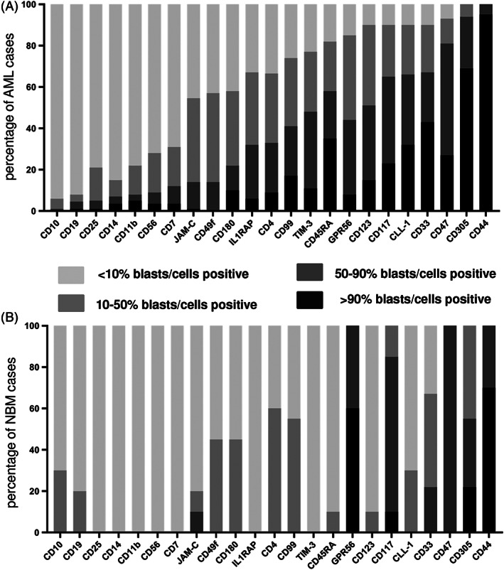
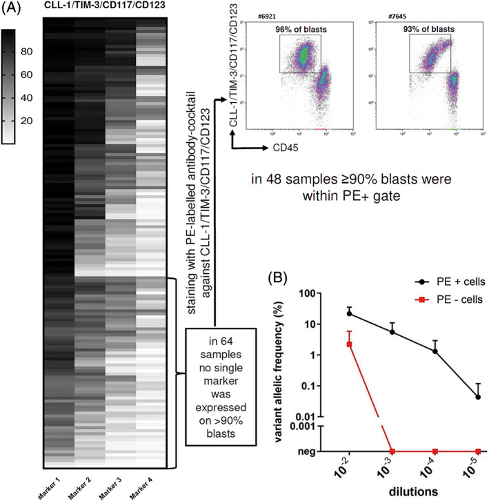
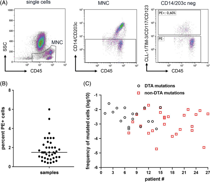
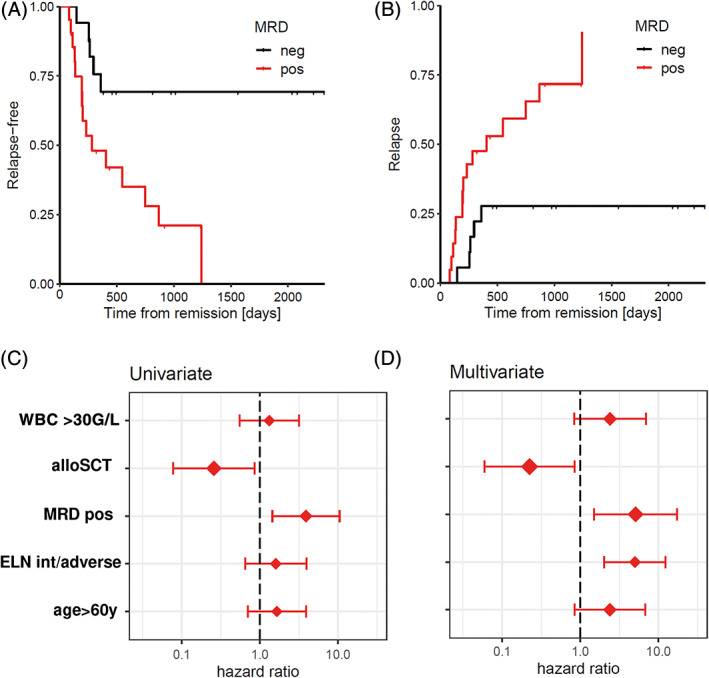
Persistent measurable residual disease (MRD) is an increasingly important prognostic marker in acute myeloid leukemia (AML). Currently, MRD is determined by multi-parameter flow cytometry (MFC) or PCR-based methods detecting leukemia-specific fusion transcripts and mutations. However, while MFC is highly operator-dependent and difficult to standardize, PCR-based methods are only available for a minority of AML patients. Here we describe a novel, highly sensitive and broadly applicable method for MRD detection by combining MFC-based leukemic cell enrichment using an optimized combinatorial antibody panel targeting CLL-1, TIM-3, CD123 and CD117, followed by mutational analysis of recurrently mutated genes in AML. In dilution experiments this method showed a sensitivity of 10-4 to 10-5 for residual disease detection. In prospectively collected remission samples this marker combination allowed for a median 67-fold cell enrichment with sufficient DNA quality for mutational analysis using next generation sequencing (NGS) or digital PCR in 39 out of 41 patients. Twenty-one samples (53.8%) tested MRD positive, whereas 18 (46.2%) were negative. With a median follow-up of 559 days, 71.4% of MRD positive (15/21) and 27.8% (5/18) of MRD negative patients relapsed (P = .007). The cumulative incidence of relapse (CIR) was higher for MRD positive patients (5-year CIR: 90.5% vs 28%, P < .001). In multivariate analysis, MRD positivity was a prominent factor for CIR. Thus, MFC-based leukemic cell enrichment using antibodies against CLL-1, TIM-3, CD123 and CD117 followed by mutational analysis allows high sensitive MRD detection and is informative on relapse risk in the majority of AML patients.


Conflict of interest statement

B.P. and A.W. report research support from Becton Dickinson BioSciences. All other authors declare no conflict of interest.

Figures

FIGURE 1
FIGURE 1
Expression of putative enrichment markers on AML bulk cells and normal HSPCs. A, The percentages of AML samples or B, CD34 + 38‐ HSPC NBM samples of which cells were either <10%, 10%–50%, 50%–90% or > 90% positive for a distinct marker, are given
FIGURE 2
FIGURE 2
Expression of enrichment markers in 150 AML samples and dilution experiment to determine the sensitivity for detecting a leukemic cell in NBM. A, Expression of enrichment markers in 150 diagnostic AML samples. The makers are arranged in descending order with the marker displaying the highest percentage of cells at the left while the marker displaying the lowest percentage of cells positive is on the right. Each row represents one patient sample. In 64 samples no single marker was expressed on >90% of blasts. Thus, these samples were analyzed with an PE‐labeled antibody cocktail targeting all four enrichment markers. Two representative examples of primary AML samples are shown. In 48 samples the majority of blasts (>90%) were then identified within the PE (marker) positive gate. B, NPM1 variant allelic frequency (VAF) of sorted PE positive (black) and PE negative cells (red) of various dilutions of three NPM1 W288fs*12 mutated leukemic samples mixed with normal BM cells (mean ± SE). neg, negative; NBM, normal bone marrow [Color figure can be viewed at wileyonlinelibrary.com]
FIGURE 3
FIGURE 3
Leukemic cell enrichment using MFC‐based sorting followed by parallel sequencing for MRD detection. A, Sorting strategy for enrichment of residual leukemic cells. The MNCs were identified using SSC low and CD45 low. The monocytes and basophils were excluded using CD14/CD203c. The CD14/CD203c negative cells were gated on marker cocktail and the marker positive and marker negative fractions were sorted. B, Percentage of sorted PE (marker) positive cells calculated as percentage of total nucleated BM cells obtained after FACS based cell sorting in 41 remission samples. C, Calculated frequencies of mutated cells detected during complete remission using our two‐step MRD detection method. Black circles denote mutated cell frequencies as detected by DTA mutations and red squares display mutated cell frequencies as detected by non‐DTA mutations. MFC, multi‐parameter flow cytometry; MRD, measurable residual disease; MNC, mononuclear cells; DTA, DNMT3A, TET2 or ASXL1 [Color figure can be viewed at wileyonlinelibrary.com]
FIGURE 4
FIGURE 4
Relapse‐free survival (RFS) and cumulative incidence of relapse (CIR) in AML patients receiving intensive chemotherapy according to MRD status. A, RFS of AML patients according to MRD status (n = 39). Patients with a positive MRD status as measured by our two‐step MRD assay had a significantly shorter duration of RFS (P = .0031). B, Competing risk analysis for CIR in AML patients according to their MRD status. C, Box plot displaying univariate analysis of hazard ratios of risk factors for CIR. D, Box plot displaying multivariate analysis of hazard ratios of risk factors for CIR. alloSCT, allogeneic stem cell transplantation; MRD pos, measureable residual disease positive; ELN int/adverse, European Leukemia Net intermediate/adverse risk [Color figure can be viewed at wileyonlinelibrary.com]

References

    1. Bachas C, Schuurhuis GJ, Assaraf YG, et al. The role of minor subpopulations within the leukemic blast compartment of AML patients at initial diagnosis in the development of relapse. Leukemia. 2012;26:1313‐1320. - PubMed
    1. Döhner H, Weisdorf DJ, Bloomfield CD. Acute myeloid leukemia. N Engl J Med. 2015;373:1136‐1152. - PubMed
    1. Yin JA, O'Brien MA, Hills RK, Daly SB, Wheatley K, Burnett AK. Minimal residual disease monitoring by quantitative RT‐PCR in core binding factor AML allows risk stratification and predicts relapse: results of the United Kingdom MRC AML‐15 trial. Blood. 2012;120:2826‐2835. - PubMed
    1. Rücker FG, Agrawal M, Corbacioglu A, et al. Measurable residual disease monitoring in acute myeloid leukemia with t(8;21)(q22;q22.1): results from the AML Study Group. Blood. 2019;134:1608‐1618. - PMC - PubMed
    1. Ivey A, Hills RK, Simpson MA, et al. Assessment of minimal residual disease in standard‐risk AML. N Engl J Med. 2016;374(5):422‐433. - PubMed