. 2020 Nov 24;14(11):14627-14634.
doi: 10.1021/acsnano.0c03125. Epub 2020 Jul 13.

Bulk Self-Assembly of Giant, Unilamellar Vesicles

Affiliations

Bulk Self-Assembly of Giant, Unilamellar Vesicles

James T Kindt et al. ACS Nano. .

Abstract

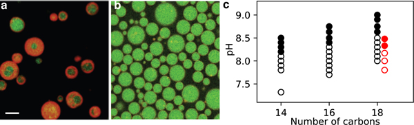
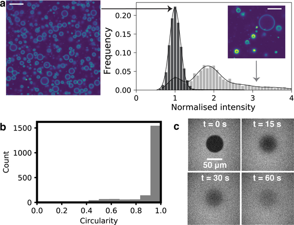
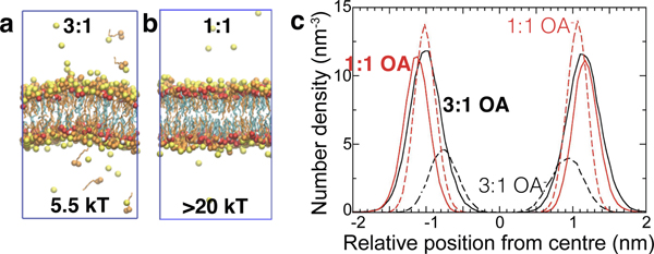
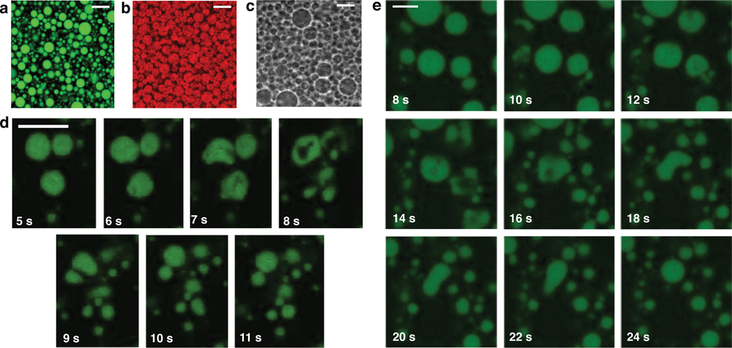
The desire to create cell-like models for fundamental science and applications has spurred extensive effort toward creating giant unilamellar vesicles (GUVs). However, a route to selectively self-assemble GUVs in bulk has remained elusive. In bulk solution, membrane-forming molecules such as phospholipids, single-tailed surfactants, and block copolymers typically self-assemble into multilamellar, onion-like structures. So although self-assembly processes can form nanoscale unilamellar vesicles, scaffolding by droplets or surfaces is required to create GUVs. Here we show that it is possible to bulk self-assemble cell-sized GUVs with almost complete selectivity over other vesicle topologies. The seemingly paradoxical pair of features that enables this appears to be having very dynamic molecules at the nanoscale that create unusually rigid membranes. The resultant self-assembly pathway enables encapsulation of molecules and colloids and can also generate model primitive cells that can grow and divide.

Keywords: giant unilamellar vesicle; liposome; membrane; origins of life; protocell; self-assembly; vesicle.


Figures

Figure 1:
Figure 1:
Fatty acid self-assembly depends on the aqueous solution conditions. a. Confocal microscopy revealed that oleic acid self-assembled into giant vesicles capable of encapsulating and retaining RNA oligomers (green). The membrane was dyed with 10 μM rhodamine B (red). The giant vesicles that self-assembled in 200 mM Na+ bicine, pH 8.43 were very heterogeneous in morphology. b. The giant vesicles that self-assembled in 50 mM Na+ bicine, pH 8.43 appeared to be unilamellar. c. The pHs at which GUVs assembled for myristoleic acid (14 carbons), palmitoleic acid (16 carbons) and oleic acid (18 carbons) in 50 mM Na+ bicine are shown as open circles (black), and the pHs at which MLVs formed are shown as solid circles. The red circles show the self-assembly of oleic acid MLVs (solid circles) and GUVs (open circles) in 250 mM Na+ bicine. The scale bar represents 10 μm.
Figure 2:
Figure 2:
Oleic acid can self-assemble into GUVs that look very static, but are in fact fluid. a. A histogram of mean membrane fluorescence intensities (see Methods) normalized against vesicles with the lowest mean membrane intensity per pixel for oligolamellar samples (grey, n = 1391) shows multiple peaks, whereas samples that appear uniform only have a single peak (black, n = 20347). Scale bar represents 10 μm. b. Oleic acid GUVs appear very spherical (see Methods). Circles have a circularity of 1. c. FRAP experiments revealed that oleic acid membranes have rapid lateral diffusion. A large bleached area (diameter 54 μm) was used to minimize the effect of diffusion during photobleaching (see also Fig. S6).
Figure 3:
Figure 3:
Self-assembly outcomes are dictated by the protonation state of the membranes. a. Molecular dynamics simulations of an octanoic acid (OA) bilayer with three octanoates for every octanoic acid molecule (3:1) showed that such bilayers exhibited short-wavelength fluctuations, and have a bending modulus Kc equal to 5.5 kT. b. Molecular dynamics simulations of an OA bilayer with one octanoate for every octanoic acid molecule (1:1) showed that such bilayers fluctuate less, and have a bending modulus Kc greater than 20 kT. c. The profiles showing distributions of oxygen sites perpendicular to the bilayer are shown for both 3:1 and 1:1 systems. The 3:1 bilayer was thinner than the 1:1 bilayer.
Figure 4:
Figure 4:
Fatty acid GUVs can encapsulate a wide range of materials. Materials encapsulated include a. pyranine (1 mM), b. 30-nm-diameter Nile Red dyed latex nanoparticles (0.1% w/v), and c. 400-nm-diameter polystyrene latex beads (0.1% w/v; see also Video S13). d–e. Oleic acid vesicles can encapsulate RNA (the same RNA oligomer as in Fig. 1a) and divide upon exposure to oleate micelles pipetted nearby. The time shown represents the number of seconds after injecting oleate micelles. Scale bar represents 10 μm for all images.

References

    1. Jung HT; Coldren B; Zasadzinski JA; Iampietro DJ; Kaler EW The Originsof Stability of Spontaneous Vesicles. Proc. Natl. Acad. Sci. U. S. A. 2001, 98, 1353–1357. - PMC - PubMed
    1. Kaler EW; Murthy AK; Rodriguez BE; Zasadzinski JA Spontaneous VesicleFormation in Aqueous Mixtures of Single-Tailed Surfactants. Science 1989, 245, 1371–1374. - PubMed
    1. Pautot S; Frisken BJ; Weitz DA Production of Unilamellar Vesicles Using anInverted Emulsion. Langmuir 2003, 19, 2870–2879.
    1. Abkarian M; Loiseau E; Massiera G. Continuous Droplet Interface Crossing Encapsulation (cDICE) for High Throughput Monodisperse Vesicle Design. Soft Matter 2011, 7, 4610.
    1. Deng N-N; Yelleswarapu M; Huck WTS Monodisperse Uni- and Multicompartment Liposomes. J. Am. Chem. Soc. 2016, 138, 7584–7591. - PubMed

Publication types