

An Avant-Garde Model of Injury-Induced Regenerative Vaginal Wound Healing
- PMID: 32602816
- PMCID: PMC7906868
- DOI: 10.1089/wound.2020.1198
An Avant-Garde Model of Injury-Induced Regenerative Vaginal Wound Healing
Abstract
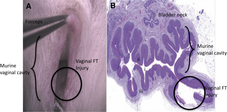
Objective: To design and validate a novel murine model of full-thickness (FT) vaginal wound healing that mirrors postinjury tissue repair and underscores the impact of estrogen signaling-driven healing kinetics, inflammation, and neovascularization. Approach: Five-week-old female CD1 mice were subjected to two 1-mm FT wounds. To assess wound healing kinetics, vaginas were harvested at 6, 12, 18, 24, 48, and 72 h and 7 days postinjury. Wounds from all time points were analyzed by hematoxylin and eosin and trichrome to, respectively, assess the rate of wound closure and tissue deposition. Inflammatory leukocyte (CD45), neutrophil (Ly6G), and macrophage (F480 and CD206) infiltration was examined by immunohistochemistry (IHC) and the resulting anti-inflammatory M2 (CD206)/total (F480) macrophage ratio quantified. Neovascularization (CD31) and estrogen receptor-α (ERα) expression levels were similarly determined by IHC. Results: We observed rapid healing with resolution of mucosal integrity by 48 h (p < 0.05), and overall neutrophils and polarized type 2 macrophages (M2) apexed at 12 h and reduced to near control levels by day 7 postinjury. Tissue repair was virtually indistinguishable from the surrounding vagina. CD31+ vessels increased between 12 h and day 7 and ERα trended to decrease at 12 h postinjury and rebound at day 7 to uninjured levels. Innovation: A proof-of-concept murine model to study vaginal wound healing kinetics and postinjury regenerative repair in the vagina was developed and verified. Conclusion: We surmise that murine vaginal mucosal repair is accelerated and potentially regulated by estrogen signaling through the ERα, thus providing a cellular and molecular foundation to understand vaginal healing responses to injury.
Keywords: estrogen receptor; novel murine injury model; regenerative wound healing; vaginal injury; wound kinetics.
Conflict of interest statement
No competing financial interests exist. No ghostwriters were enlisted.
Figures


References
-
- Vaginal Agenesis. USNational Libr Med. https://ghr.nlm.nih.gov/condition/mayer-rokitansky-kuster-hauser-syndrom... (accessed July31, 2019)
-
- Androgen Insensitivity Syndrome. USNational Libr Med 2017 [cited Oct 2, 2017]. https://ghr.nlm.nih.gov/condition/androgen-insensitivity-syndrome (accessed July31, 2019)
-
- Mayer-Rokitansky-Küster-Hauser syndrome. USNational Libr Med 2017 [cited Oct 2, 2017]. https://ghr.nlm.nih.gov/condition/mayer-rokitansky-kuster-hauser-syndrome (accessed July31, 2019)
-
- Raya-Rivera A, Esquiliano D, Fierro-Pastrana R, et al. Tissue-engineered autologous vaginal organs in patients: a pilot cohort study. Lancet 2014;384:329–336 - PubMed
-
- Godbole P, Koyle M, Wilcox D, eds. Pediatric Urology: Surgical Complications and Management. Hoboken, NJ: Second Edi Wiley Blackwell, 2015
Publication types
MeSH terms
Substances
Grants and funding
LinkOut - more resources
Full Text Sources
Other Literature Sources
Medical
Research Materials
Miscellaneous
