. 2020 Jul;68(7):491-513.
doi: 10.1369/0022155420939833. Epub 2020 Jun 30.

Immunocytochemical Localization of Olfactory-signaling Molecules in Human and Rat Spermatozoa

Affiliations

Immunocytochemical Localization of Olfactory-signaling Molecules in Human and Rat Spermatozoa

Yuliya Makeyeva et al. J Histochem Cytochem. 2020 Jul.

Abstract

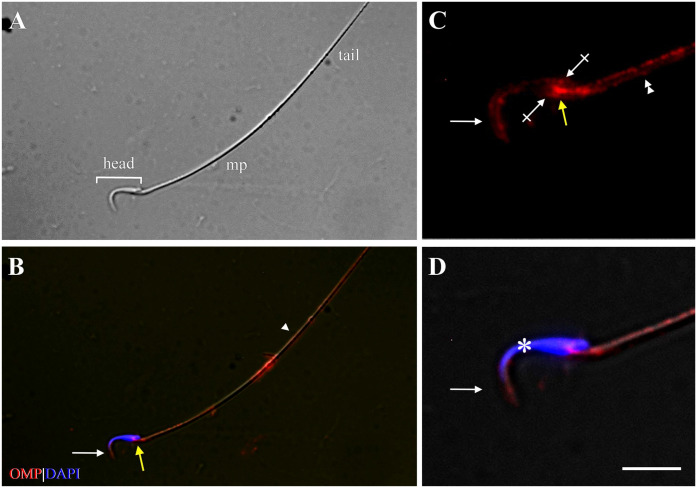
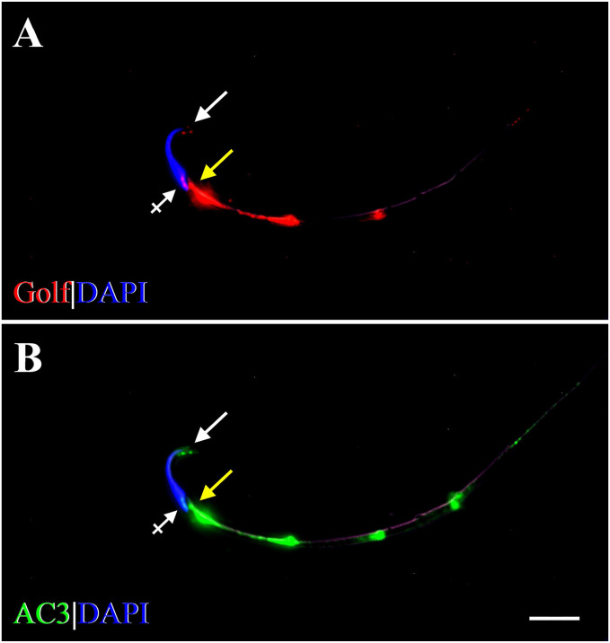
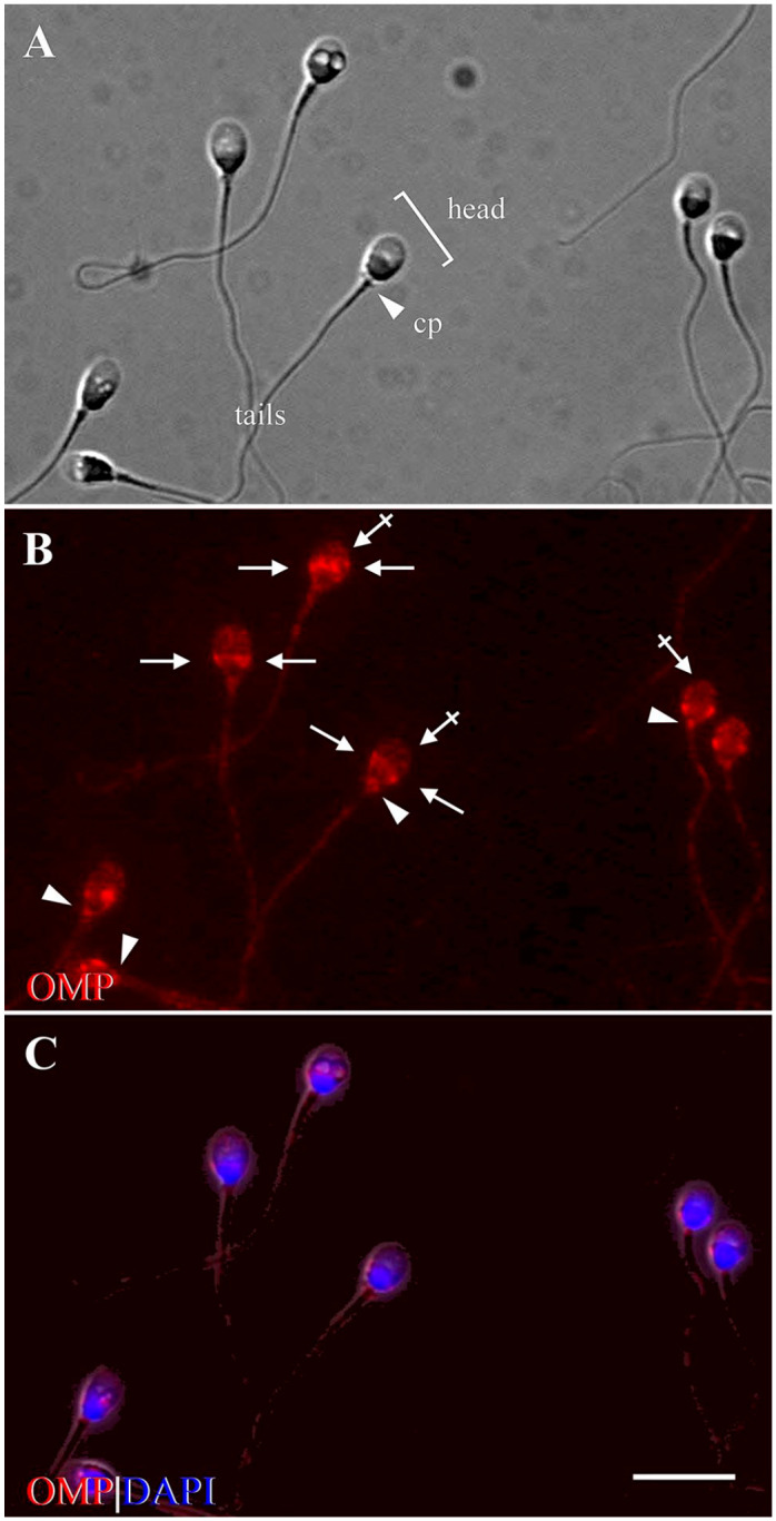
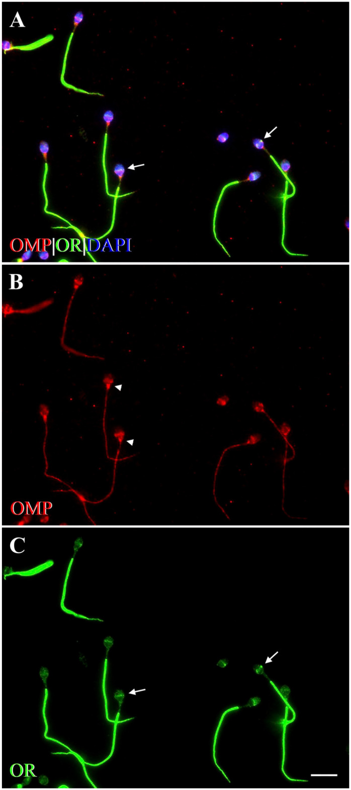
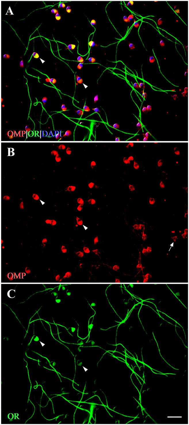
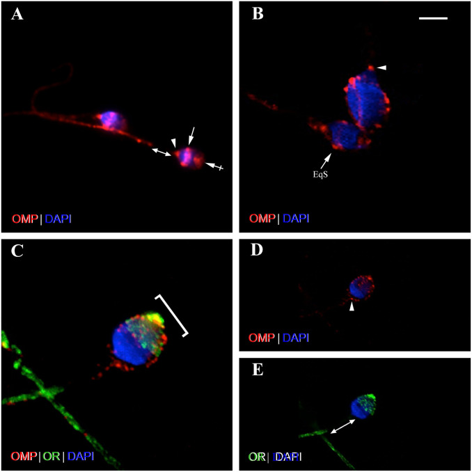
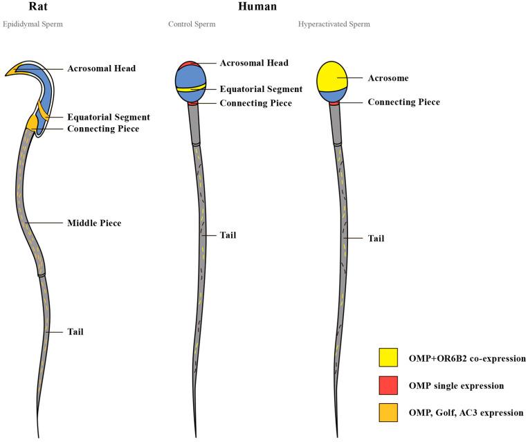
Expression of olfactory receptors (ORs) in non-olfactory tissues has been widely reported over the last 20 years. Olfactory marker protein (OMP) is highly expressed in mature olfactory sensory neurons (mOSNs) of the olfactory epithelium. It is involved in the olfactory signal transduction pathway, which is mediated by well-conserved components, including ORs, olfactory G protein (Golf), and adenylyl cyclase 3 (AC3). OMP is widely expressed in non-olfactory tissues with an apparent preference for motile cells. We hypothesized that OMP is expressed in compartment-specific locations and co-localize with an OR, Golf, and AC3 in rat epididymal and human-ejaculated spermatozoa. We used immunocytochemistry to examine the expression patterns of OMP and OR6B2 (human OR, served as positive olfactory control) in experimentally induced modes of activation and determine whether there are any observable differences in proteins expression during the post-ejaculatory stages of spermatozoal functional maturation. We found that OMP was expressed in compartment-specific locations in human and rat spermatozoa. OMP was co-expressed with Golf and AC3 in rat spermatozoa and with OR6B2 in all three modes of activation (control, activated, and hyperactivated), and the mode of activation changed the co-expression pattern in acrosomal-reacted human spermatozoa. These observations suggest that OMP expression is a reliable indicator of OR-mediated chemoreception, may be used to identify ectopically expressed ORs, and could participate in second messenger signaling cascades that mediate fertility.

Keywords: chemoreception; fluorescence microscopy; interstitial tissue; olfactory epithelium; olfactory marker protein; reproduction; sperm.


Conflict of interest statement

Competing Interests: The author(s) declared no potential conflicts of interest with respect to the research, authorship, and/or publication of this article.

Figures

Figure 1.
Figure 1.
Bright-field photomicrograph of OMP distribution in the rat OE as revealed by immunohistochemistry. The OMP brown reaction product is distributed in the somata of mature olfactory sensory neurons, in their dendrites and knobs, the cilia, and their axons. Sustentacular cells, basal cells, and immature OSNs are unlabeled. This pattern is as previously reported,, and thus serves as positive control for other rat tissues. Scale bar = 20 µm. Abbreviations: OMP, olfactory marker protein; OE, olfactory epithelium; OSNs, olfactory sensory neurons.
Figure 2.
Figure 2.
Bright-field photomicrograph of olfactory marker protein (OMP) staining in the rat testes. (A) Control tissue containing seminiferous tubules (ST) and interstitial tissue (IT) is stained by cresyl violet but there is no immunostaining when the OMP antibody is left out. Leydig-like cells are shown on the right at higher magnification and are void of OMP reaction product. (B) Tissue stained by OMP antibodies reveal Leydig-like cells express OMP (brown) and these are only found in the IT. OMP+ Leydig-like cells are shown on the right at higher magnification. Scale bar = 50 µm for the tissue and 18 µm for the panel of individual cells.
Figure 3.
Figure 3.
Identification of OMP+ staining of Leydig cells in the rat testes. (A) Photomicrograph of calretinin (CR) immunostaining (green) of cells of IT. CR is a marker for Leydig cells., The somata of Leydig cells are polygonal or round-oval in shape and have positively stained cytoplasm and prominent round nuclei. (B) Photomicrograph shows Leydig cells stained with OMP (red). OMP immunoreactivity varies in intensity between individual OMP+ cells but no OMP+ staining is observed in cells of the ST. (C) OMP is co-expressed with CR using double-labeling IHC as revealed by yellow-orange cytoplasm in merged image. CR immunoreactivity can be observed singly in some IT cells but all OMP+-stained cells are co-stained with CR. DAPI (blue) stains cell nuclei. Scale bar = 20 µm. Abbreviations: OMP, olfactory marker protein; IT, interstitial tissue; ST, seminiferous tubule; IHC, immunohistochemistry; DAPI, 4′,6-diamidino-2-phenylindole dihydrochloride.
Figure 4.
Figure 4.
Graph of Leydig and OMP+ cells representation in the IT of the rat testes. (A) Leydig cell population, as defined by immunolabeling by CR (CR+), represent the majority—79% of all cells (n=115) in the IT of rat testes. The CR-negative (CR–) cells represent the remaining 21%. (B) 81% of the Leydig cell population (n=91) was OMP+ (Double: CR+OMP+). No cells are labeled by OMP alone (OMP Single+) and CR+ only cells (CR Single+) are represented 19% of the Leydig cell population. Statistical significance between groups is indicated by asterisks. CR+ vs CR– (χ2 test, *p<0.001); double vs CR single+ (χ2 test, *p<0.001); double vs OMP single+ (F test, *p<0.001). Abbreviations: OMP, olfactory marker protein CR, calretinin; IT, interstitial tissue.
Figure 5.
Figure 5.
Expression of OMP showing compartment-specific distribution in the rat spermatozoa. (A) Phase-contrast photomicrograph shows the typical structure of rat spermatozoa: head (bracket), mp, and tail. (B) Immunofluorescence image shows expression of OMP (red) on the head (white arrow), cp (yellow arrow), and tail of spermatozoa, with strongest expression along the mp. The arrowhead marks the border of the mp and the tail. (C) High-power photomicrograph shows OMP expression on the acrosomal region (arrow) and EqS (crossed arrows) of the head, the cp (yellow arrow), and the mp (double arrowhead). (D) DAPI (blue) stains the spermatozoa nucleus (asterisk). Scale bar = 20 µm for low-power photomicrographs (A and B) and 8 µm for high-power photomicrographs of head region (C and D). Abbreviations: OMP, olfactory marker protein; mp, middle piece; cp, connecting piece; EqS, equatorial segment; DAPI, 4′,6-diamidino-2-phenylindole dihydrochloride.
Figure 6.
Figure 6.
Fluorescent photomicrographs showing compartment-specific expression of Golf and AC3 in the rat spermatozoa. (A) Expression of Golf (red) is seen on the acrosomal region (arrow) and EqS (crossed arrow) of the head, on the cp (yellow arrow), and along the tail in a discontinuous pattern. (B) Expression of AC3 (green) is seen in similar compartments: on the head (arrow, crossed arrow), on the cp (yellow arrow), and along the tail in a discontinuous pattern. Strong expression of Golf and AC3 is seen on the mp, gradually diminishing along the tail. DAPI (blue) stains spermatozoal nuclei. Scale bar = 10µm. Abbreviations: AC3, adenylyl cyclase 3; Golf, olfactory G protein; EqS, equatorial segment; cp, connecting piece; mp, middle piece; DAPI, 4′,6-diamidino-2-phenylindole dihydrochloride.
Figure 7.
Figure 7.
Photomicrographs show compartment-specific expression of OMP in ejaculated human spermatozoa (control). (A) Phase-contrast image shows the structure of human spermatozoa, consisting of the head (bracket), cp (arrowhead), and tail. (B) Immunofluorescence shows expression of OMP (red) on the apical part of the acrosomal region (crossed arrows) and along the EqS of the head (arrows), on the cp (arrowheads), and along the tail, except for the mp. (C) DAPI (blue) shows spermatozoal nuclei, surrounded by a thin rim of cytoplasm. Acrosome is seen above nuclei, on the anterior part of the head (crossed arrows in B). Scale bar = 10 µm. Abbreviations: OMP, olfactory marker protein; cp, connecting piece; EqS, equatorial segment; mp, middle piece; DAPI, 4′,6-diamidino-2-phenylindole dihydrochloride.
Figure 8.
Figure 8.
Fluorescent photomicrographs showing co-expression of OMP with OR6B2 in control human spermatozoa. OR6B2, a marker for a human OR, was co-labeled with OMP using double-labeling ICC. (A) OMP co-expresses with OR6B2 (yellow) on the EqS (arrows) of the head, and along the tail, except for the mp. DAPI (blue) stains spermatozoal nuclei. (B) Immunofluorescence shows expression of OMP (red) on the head, cp (arrowheads), and along the tail. On the head, expression of OMP is seen prominently on the EqS and faintly on the apical part of acrosomal region. OMP is also expressed in a discontinuous and punctate pattern along the tail, except for the mp. (C) Expression of OR6B2 (OR, green) is seen on the EqS of the head (arrows) and evenly along the tail, but not on the cp or mp. Scale bar = 10µm. Abbreviations: OMP, olfactory marker protein; OR, olfactory receptor (OR6B2); ICC, immunocytochemistry; EqS, equatorial segment; DAPI, 4′,6-diamidino-2-phenylindole dihydrochloride; cp, connecting piece; mp, middle piece.
Figure 9.
Figure 9.
Fluorescent photomicrographs showing co-expression of OMP with OR6B2 in activated human spermatozoa using double-labeling ICC. (A) OMP co-expresses with OR6B2 (yellow) on the head and tail, but not on the mp. On the head, prominent co-expression is seen across the entire acrosomal region (arrowheads). DAPI (blue) stains spermatozoal nuclei. (B) Expression of OMP (red) is observed on the head, the cp, and along the tail. In some spermatozoa, OMP expression is seen across the entire acrosomal region (arrowheads). In other spermatozoa, OMP expression is seen primarily on the EqS (arrow). OMP is expressed along the tail in a discontinuous pattern. (C) Expression of OR6B2 (OR, green) is seen on the head (arrowheads), and tail in some spermatozoa, whereas in other spermatozoa, OR6B2 expression is seen only along the tail. No single expression of OMP or OR6B2 is seen on the mp. The pattern of staining in activated spermatozoa is distinct from that of control (unactivated) spermatozoa. Scale bar = 10µm. Abbreviations: OMP, olfactory marker protein; OR, olfactory receptor (OR6B2); ICC, immunocytochemistry; mp, middle piece; DAPI, 4′,6-diamidino-2-phenylindole dihydrochloride; cp, connecting piece; EqS, equatorial segment.
Figure 10.
Figure 10.
Photomicrographs show confocal Z stack images of human spermatozoa in control (A and B) and activated (C to E) modes. Activation of spermatozoa affects OMP and OR6B2 expression pattern on the head. Photomicrographs (A and B) show control (unactivated) spermatozoa stained with OMP using single-labeling ICC. (A) Similar patterns of OMP expression (red) are seen in two control spermatozoa. OMP expression is seen on the EqS (arrow) and on the apical part of the acrosomal region (crossed arrow) of the head, on the cp (arrowhead), and, in a discontinuous pattern, along entire tail. No OMP expression is seen on the mp (double head arrow). (B) Photomicrograph shows heads of two control spermatozoa in two rotations. Dot-like expression of OMP (red) is seen on the EqS (arrow) and on the apical part of the acrosomal region of the head, and on the cp (arrowhead). Photomicrographs (C to E) show activated spermatozoon co-stained with OMP and OR6B2 using double-labeling ICC. Representative co-expression is shown in merged image C, and representative expression of each antibody is displayed in image D (OMP) and image E (OR6B2). (C) Three-dimensional image stack reconstruction of spermatozoon shows co-expression of OMP with OR6B2 (yellow) in dot-like patterns across the entire acrosomal region of the head (bracket), and along the tail. On the head, strongest co-expression is visible on the apical tip. (D) Single-image slice shows OMP (red) expression across the entire acrosomal region of the head in defined dot-like pattern, and along the tail. OMP expression is seen on the cp (arrowhead), and a few spots of OMP staining are seen in near proximity. (E) Single-image slice shows OR6B2 (OR, green) expression across the entire acrosomal region of the head in a dot-like pattern, and along the tail. No OR6B2 expression is visible on the cp or mp (double head arrow). DAPI (blue) stains spermatozoa nuclei. Scale bar = 5 µm for B, C and 10µm for A, D, E. Abbreviations: OMP, olfactory marker protein; OR, olfactory receptor (OR6B2); ICC, immunocytochemistry; EqS, equatorial segment; cp, connecting piece; mp, middle piece; DAPI, 4′,6-diamidino-2-phenylindole dihydrochloride.
Figure 11.
Figure 11.
OMP, OR6B2, Golf, and AC3 are localized in compartment-specific locations in human and rat spermatozoa. In rat spermatozoa, OMP, Golf, and AC3 are simultaneously expressed on the head (acrosomal region and equatorial segment), the connecting piece, and along the tail, with strongest expression on the middle piece. In control human spermatozoa, OMP co-expresses with OR6B2 on the head (equatorial segment), and along the tail, except for the middle piece. In the hyperactivated spermatozoa, OMP co-expresses with OR6B2 on the head (acrosome), and along the tail, except for the middle piece. In control and hyperactivated spermatozoa, OMP is singly expressed on the connecting piece. A clear difference in the pattern of OMP expression between human (both control and hyperactivated) and rat spermatozoa is observed on the middle piece. The differences in the pattern of OMP and OR6B2 co-expression between the control and hyperactivated human spermatozoa are observed on the head, with no changes of OMP single expression on the connecting piece. Abbreviations: OMP, olfactory marker protein; OR, olfactory receptor (OR6B2); AC3, adenylyl cyclase 3; Golf, olfactory G protein.

Similar articles

Cited by

References

    1. Buck L, Axel R. A novel multigene family may encode odorant receptors: a molecular basis for odor recognition. Cell. 1991;65(1):175–87. - PubMed
    1. Shepherd G. Discrimination of molecular signals by the olfactory receptor neuron. Neuron. 1994;13(4):771–90. - PubMed
    1. Giorgi F, Maggio R, Bruni L. Are olfactory receptors really olfactive? Biosemiotics. 2011;4(3):331–47.
    1. Buck L. Information coding in the vertebrate olfactory system. Annu Rev Neurosci. 1996;19(1):517–44. - PubMed
    1. Malnic B, Hirono J, Sato T, Buck L. Combinatorial receptor codes for odors. Cell. 1999;96(5):713–23. - PubMed

Publication types

Substances