. 2020 Aug 6;5(15):e136706.
doi: 10.1172/jci.insight.136706.

Erythromycin inhibits neutrophilic inflammation and mucosal disease by upregulating DEL-1

Affiliations

Erythromycin inhibits neutrophilic inflammation and mucosal disease by upregulating DEL-1

Tomoki Maekawa et al. JCI Insight. .

Abstract

Macrolide antibiotics exert antiinflammatory effects; however, little is known regarding their immunomodulatory mechanisms. In this study, using 2 distinct mouse models of mucosal inflammatory disease (LPS-induced acute lung injury and ligature-induced periodontitis), we demonstrated that the antiinflammatory action of erythromycin (ERM) is mediated through upregulation of the secreted homeostatic protein developmental endothelial locus-1 (DEL-1). Consistent with the anti-neutrophil recruitment action of endothelial cell-derived DEL-1, ERM inhibited neutrophil infiltration in the lungs and the periodontium in a DEL-1-dependent manner. Whereas ERM (but not other antibiotics, such as josamycin and penicillin) protected against lethal pulmonary inflammation and inflammatory periodontal bone loss, these protective effects of ERM were abolished in Del1-deficient mice. By interacting with the growth hormone secretagogue receptor and activating JAK2 in human lung microvascular endothelial cells, ERM induced DEL-1 transcription that was mediated by MAPK p38 and was CCAAT/enhancer binding protein-β dependent. Moreover, ERM reversed IL-17-induced inhibition of DEL-1 transcription, in a manner that was dependent not only on JAK2 but also on PI3K/AKT signaling. Because DEL-1 levels are severely reduced in inflammatory conditions and with aging, the ability of ERM to upregulate DEL-1 may lead to a novel approach for the treatment of inflammatory and aging-related diseases.

Keywords: Immunology; Inflammation; Mouse models; Neutrophils; endothelial cells.


Conflict of interest statement

Conflict of interest: The authors have declared that no conflict of interest exists.

Figures

Figure 1
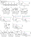
Figure 1. ERM upregulates DEL-1 mRNA and protein levels.
(A) DEL1 mRNA transcription was analyzed by quantitative PCR (qPCR) in HMVECs incubated for the indicated time periods with or without ERM (10 μg/mL), JSM (10 μg/mL), or PC (10 μg/mL). Data were normalized against GAPDH mRNA and expressed as fold induction relative to treatment with ethanol (vehicle control), which was assigned an average value of 1. (B) DEL-1 protein levels in the cell culture supernatants after 6-hour incubation were measured by ELISA. (C) Del1 mRNA transcription in the lung tissue was analyzed by qPCR 24 hours after i.p. injection of ethanol (vehicle control), ERM (20 mg/kg), JSM (20 mg/kg), or PC (20 mg/kg). Data were normalized against Gapdh mRNA and expressed as fold induction relative to treatment with ethanol (vehicle control), which was assigned an average value of 1. Data are presented as the mean ± SD; (A and B: n = 6 sets of HMVECs cultures; and C: n = 6 mice/group); (A) 2-way ANOVA followed by Holm-Šidák multiple comparisons test; (B and C) 1-way ANOVA followed by Tukey’s multiple comparisons test; *P < 0.01, ***P < 0.0001 between indicated groups.
Figure 2
Figure 2. ERM suppresses neutrophil infiltration in BALF.
(A) Experimental design. E. coli LPS (2.5 mg/kg) was administrated intratracheally. ERM (20 mg/kg) or JSM (20 mg/kg), PC (20 mg/kg), or ethanol control (n = 10 mice/group) was administrated i.p. 3 hours before and 24 hours after LPS administration. Samples were collected 48 hours after LPS administration. (B and C) Neutrophil counts (B) and myeloperoxidase (MPO) activity (C) in the BALF 48 hours after LPS challenge (B: n = 10 mice/group; C: n = 6 mice/group). (D) The mRNA levels of proinflammatory cytokines (Il6, Il17, and Tnf), Del1, and Il10 in the lung tissue were determined by qPCR 48 hours after LPS challenge (n = 6 mice/group). Data were normalized against Gapdh mRNA and expressed as fold induction relative to treatment with ethanol control, which was assigned an average value of 1. (E) Mean linear intercept measured in central and peripheral areas of the lungs 48 hours after LPS challenge (n = 10 mice/group). (F) Representative images of H&E-stained pulmonary parenchyma 48 hours after LPS challenge. Upper panel: scale bars, 50 μm; lower panel: scale bars, 25 μm. (G) IHC of lung tissue in WT mice stained with DEL-1 and neutrophil elastase 48 hours after LPS challenge. Scale bars: 50 μm. (H) IHC of lung tissue in Del1–/– mice stained with DEL-1 and neutrophil elastase 48 hours after sublethal LPS (2.5 mg/kg) challenge as outlined in panel A. Scale bars: 100 μm. Data are presented as the mean ± SD. **P < 0.01, ***P < 0.001 by 1-way ANOVA followed by Tukey’s multiple comparisons test.
Figure 3
Figure 3. ERM improves mouse survival after LPS-induced acute lung injury in a manner comparable to that of DEL-1–Fc.
(A) Experimental design. E. coli LPS (25 mg/kg; lethal dose) was administrated intratracheally. ERM, JSM, PC (all 3 antibiotics at 20 mg/kg), or ethanol control was administrated i.p., while DEL-1–Fc (10 μg) or Fc control (3.3 μg; equal molar amount with 10 μg DEL-1–Fc) was administered intravenously, at the indicated time points. (B) Survival rates for mice treated with ethanol control, ERM, JSM, or PC and subjected to acute lung injury by LPS (n = 14 mice/group). (CE) Determination of TNF (C), IL-17 (D), and IL-10 (E) serum levels in LPS-challenged mice treated with ERM (or controls) or DEL-1–Fc (or Fc control); serum was collected 24 hours after LPS administration (n = 6 mice/group). (F) Survival rates of mice treated with DEL-1–Fc or Fc-control and subjected to acute lung injury by LPS (n = 14 mice/group). (G) Dynamics of oxygen saturation (SpO2) levels in mice subjected to LPS-induced acute lung injury over the course of 24 hours following LPS administration and treatment with the indicated antibiotics (left panel), DEL-1–Fc (right panel), or controls (ethanol or Fc) (n = 10 mice/group). (H and I) WT and Del1–/– mice were challenged with LPS and treated with ERM (or ethanol control) or DEL-1–Fc (or Fc control) as outlined in panel A. Neutrophil numbers were calculated in the BALF of WT and Del1–/– mice 24 hours after LPS administration (n = 10 mice/group) (H). Survival rate of WT and Del1–/– mice subjected to LPS-induced acute lung injury (n = 14 mice/group) (I). (JL) Serum levels of TNF (J), IL-17 (K), and IL-10 (L) in LPS-challenged WT and Del1–/– mice treated with ERM (or ethanol control) or DEL-1–Fc (or Fc control); serum was collected 24 hours after LPS administration (n = 6 mice/group). (M) Survival rate of LPS-challenged Del1–/– mice treated with ERM (or ethanol control) or DEL-1–Fc (or Fc control) (n = 14 mice/group). Data are presented as the mean ± SD. **P < 0.01 by the log-rank test (B, F, I, and M). **P < 0.01, ***P < 0.001 by 1-way ANOVA followed by Tukey’s multiple comparisons test (C, D, E, H, J, K, and L). ***P < 0.001 by 2-way ANOVA followed by Holm-Šidák multiple comparisons test (G).
Figure 4
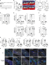
Figure 4. ERM suppresses ligature-induced inflammatory bone loss in a DEL-1–dependent manner.
(A) Experimental design. Periodontal bone loss was induced in WT or Del1–/– mice for 9 days by ligating a maxillary second molar and leaving the contralateral tooth unligated (baseline control). Groups of mice were given ERM (20 mg/kg), JSM (20 mg/kg), PC (20 mg/kg), or ethanol control i.p. every day until the day before sacrifice (day 8). (B) Measurements of bone loss in the indicated groups of LIP-subjected mice (left panel; n = 10 mice/group) and representative images of maxillae from each group (right panel). (C) Bone loss was measured in littermate WT or Del1–/– mice that were subjected to LIP and treated with ERM (20 mg/kg) or ethanol control as shown in panel A (n = 10 mice/group). (D) Numbers of neutrophils in the gingiva of LIP-subjected WT mice treated with ethanol control, ERM (20 mg/kg), JSM (20 mg/kg), or PC (20 mg/kg) as described above (n = 6 mice/group). (E) Relative mRNA expression of the indicated molecules in the gingival tissue from LIP-subjected WT mice treated with ERM, JSM, PC, or ethanol control as above. Data were normalized to Gapdh mRNA and are presented as fold change relative to baseline (unligated control), which was set as 1 (n = 6 mice/group). (F) Numbers of neutrophils in the gingival tissue of LIP-subjected WT or Del1–/– mice treated with ERM (20 mg/kg) or ethanol control (n = 6 mice/group). (G) Determination of the protein and mRNA levels of IL-17, IL-6, and IL-10 in the gingival tissue of LIP-subjected WT or Del1–/– mice, which were treated (or not; ethanol control) with 20 mg/kg ERM as outlined in panel A. Protein concentrations (pg cytokines/mg total protein in tissue lysates are shown) and mRNA expression were determined by ELISA and qPCR, respectively. The mRNA data were normalized to Gapdh mRNA and are presented as fold change relative to vehicle-treated WT mice, which was set as 1 (n = 6 mice/group). (H) Tissue sections from LIP-subjected WT mice were stained for DEL-1, neutrophil elastase and nuclei using DAPI. Scale bars, 100 μm. Data are presented as the mean ± SD. *P < 0.01, ***P < 0.0001 by 1-way ANOVA followed by Tukey’s multiple comparisons test (BG).
Figure 5
Figure 5. ERM reverses IL-17–mediated suppression of DEL-1 by regulating the C/EBPβ transcription factor.
(A) HMVECs were stimulated as indicated for 4 hours in the absence or presence of IL-17 (5 ng/mL). Prior to IL-17 stimulation, the cells were pretreated for 30 minutes with ERM (10 μg/mL), JSM (10 μg/mL), or PC (10 μg/mL). DEL1 mRNA expression was determined by qPCR, and data were normalized against GAPDH mRNA and expressed as fold induction relative to ethanol treatment (vehicle control), which was assigned an average value of 1 (n = 6 sets of cultures/group). (B and C) HMVECs were transiently transfected with hEDIL3-promoter-Luc reporter plasmid, pretreated for 30 minutes with or without ERM (10 μg/mL), JSM (10 μg/mL), or PC (10 μg/mL), followed by 8-hour stimulation with or without IL-17 (5 ng/mL), and analyzed for luciferase activity. A Renilla luciferase construct was cotransfected as an internal control for normalization. Data are presented as fold change relative to ethanol control treatment, which was set as 1 (n = 6 sets of cultures/group). (D) ChIP analysis of C/EBPβ occupancy at the DEL1 promoter in HMVECs treated for 4 hours with ethanol control, ERM (10 μg/m), JSM (10 μg/m), or PC (10 μg/m) (n = 4 sets of cultures/group). (E) Same experimental setup as in panel D, with included stimulation with IL-17 (5 ng/mL) for 4 hours following 30 minutes’ pretreatment with ERM and controls (n = 4 sets of cultures/group). Nonimmunoprecipitated cell extracts were used as input samples. In the experiments whose results are shown in A, C, and E, sequential treatments were performed without intermediate washing steps. Data are expressed as percentage of input. Data are presented as the mean ± SD. **P < 0.001, ***P < 0.0001 by 1-way ANOVA followed by Tukey’s multiple comparisons test.
Figure 6
Figure 6. ERM activates GHSR/JAK2 signaling for regulating DEL-1 expression.
(A) DEL1 mRNA expression determined by qPCR and DEL-1 protein levels determined by ELISA in control or GHSR siRNA-transfected HMVECs treated with ERM (10 μg/mL) or ghrelin (5 μg/mL) 3 hours (mRNA) or 6 hours (protein) (n = 6 culture sets/group). Data normalized against GAPDH mRNA are expressed as fold induction relative to ethanol (set as 1). (B) HMVECs, pretreated for 24 hours with control or GHSR siRNA (20 nM), were incubated with ERM and assayed for phosphorylation at indicated points. (C) After 1-hour pretreatment with AG490 (10 μM), LY294002 (20 μM), or SB203580 (10 μM), HMVECs were incubated 3 hours with ERM and assayed for DEL1 expression (n = 6 culture sets/group). Data normalized against GAPDH mRNA were expressed as fold induction relative to ethanol control (set as 1). (D) HMVECs were transiently transfected with hEDIL3-promoter-Luc reporter plasmid, pretreated 1 hour with inhibitors, and subsequently incubated 8 hours with ERM or control followed by luciferase assay. Data are presented as fold change relative to ethanol control, set as 1 (n = 6 culture sets/group). (E) HMVECs, pretreated as above with inhibitors, were incubated 4 hours with ERM and subjected to ChIP analysis of C/EBPβ occupancy at the EDIL3 promoter (n = 4 culture sets/group). (F) After 30-minute pretreatment with ERM or RvD1 (100 nM), HMVECs were stimulated (3 hours), or not, with IL-17 (5 ng/mL). DEL1 mRNA expression was assayed and presented as above (n = 6 HMVEC culture sets/group). (G) HMVECs were transiently transfected with hEDIL3-promoter-Luc reporter plasmid and pretreated with inhibitors. After 1 hour, the cells were treated with ERM, RvD1, or control for 30 minutes, followed by 8-hour stimulation with IL-17 and luciferase activity assay (n = 6 culture sets/group). Data are presented as fold change relative to ethanol (set as 1). (H) After 1-hour pretreatment with inhibitors, HMVECs were treated with ERM, RvD1, or ethanol for 30 minutes, followed by 4-hour stimulation with IL-17. Chromatin was immunoprecipitated with anti–C/EBPβ IgG and subjected to qPCR of the DEL1 promoter. Nonimmunoprecipitated cell extracts served as input samples. (I) After 1-hour pretreatment with inhibitors, HMVECs were incubated with RvD1 or ERM for 30 minutes and assayed for phosphorylation. In experiments shown in AI, sequential treatments were performed without intermediate washing steps. Each compound was used at the same concentration in all experiments. Data are shown as means ± SD. **P < 0.001, ***P < 0.0001 by 1-way ANOVA with Tukey’s multiple comparisons test (A and CH).

Similar articles

Cited by

References

    1. Kanoh S, Rubin BK. Mechanisms of action and clinical application of macrolides as immunomodulatory medications. Clin Microbiol Rev. 2010;23(3):590–615. doi: 10.1128/CMR.00078-09. - DOI - PMC - PubMed
    1. Seemungal TA, Wilkinson TM, Hurst JR, Perera WR, Sapsford RJ, Wedzicha JA. Long-term erythromycin therapy is associated with decreased chronic obstructive pulmonary disease exacerbations. Am J Respir Crit Care Med. 2008;178(11):1139–1147. doi: 10.1164/rccm.200801-145OC. - DOI - PubMed
    1. Domon H, et al. Mechanism of macrolide-induced inhibition of pneumolysin release involves impairment of autolysin release in macrolide-resistant Streptococcus pneumoniae. Antimicrob Agents Chemother. 2018;62(11):e00161-18. - PMC - PubMed
    1. Southern KW, Barker PM, Solis-Moya A, Patel L. Macrolide antibiotics for cystic fibrosis. Cochrane Database Syst Rev. 2012;11:CD002203. - PMC - PubMed
    1. Albert RK, et al. Azithromycin for prevention of exacerbations of COPD. N Engl J Med. 2011;365(8):689–698. doi: 10.1056/NEJMoa1104623. - DOI - PMC - PubMed

Publication types

MeSH terms