. 2020 Oct 1;130(10):5313-5325.
doi: 10.1172/JCI133310.

Everolimus improves the efficacy of dasatinib in PDGFRα-driven glioma

Affiliations

Everolimus improves the efficacy of dasatinib in PDGFRα-driven glioma

Zachary Miklja et al. J Clin Invest. .

Abstract

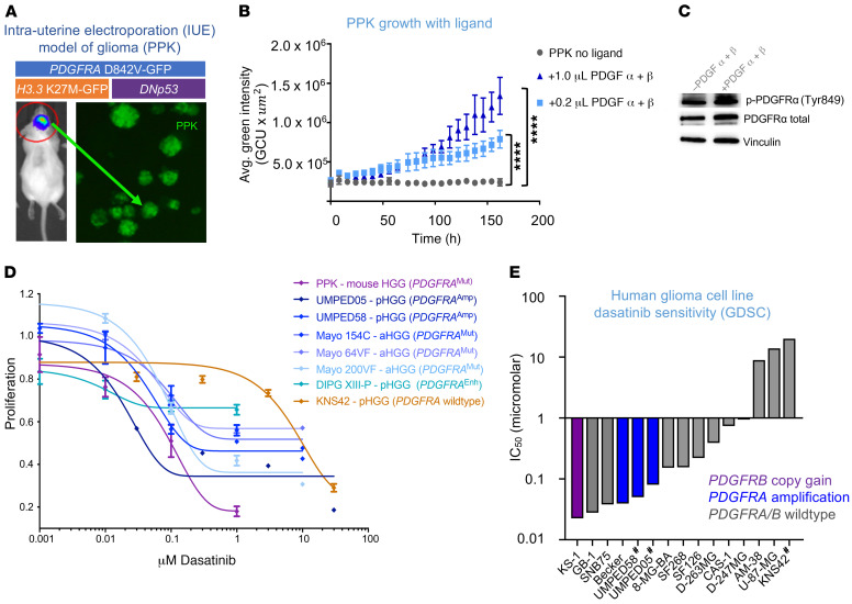
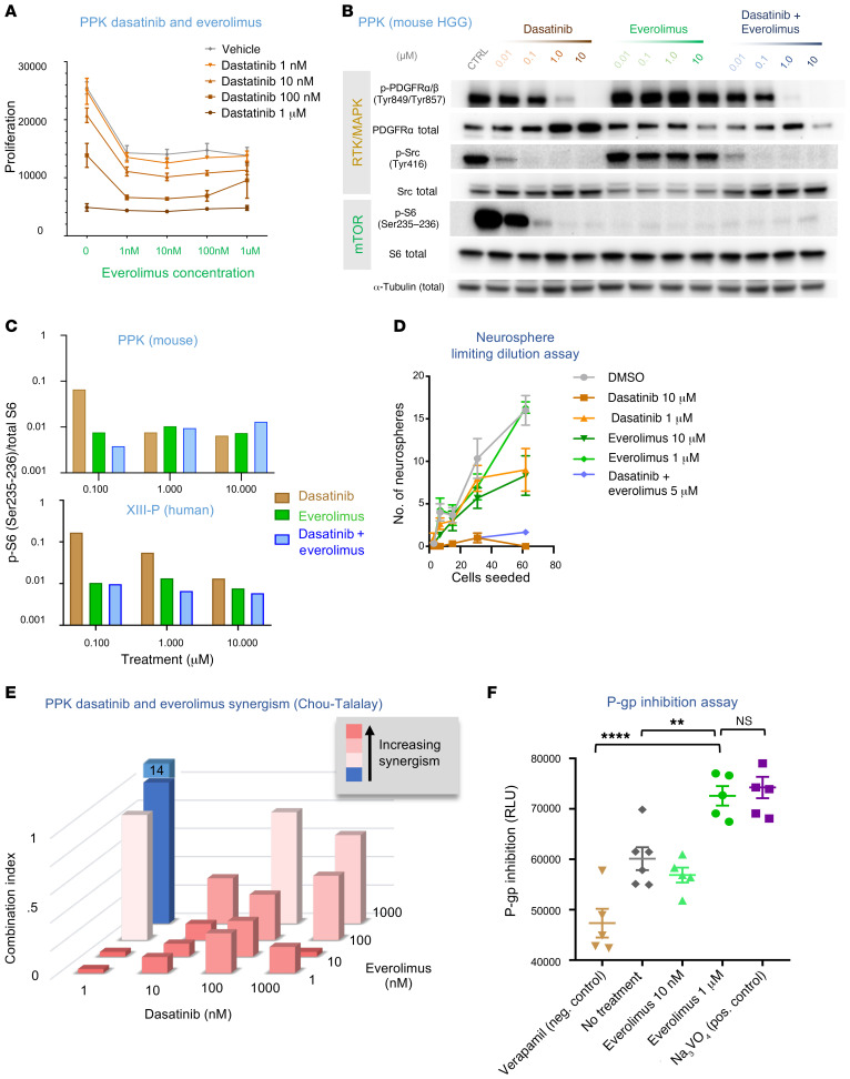
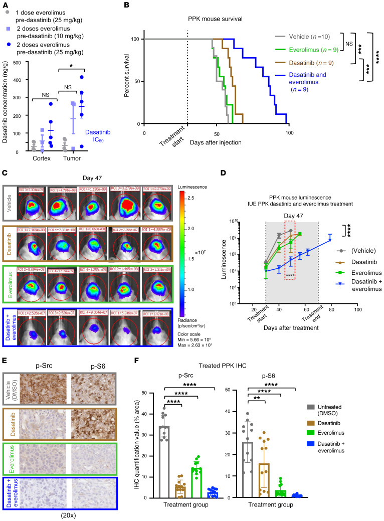
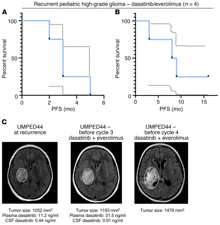
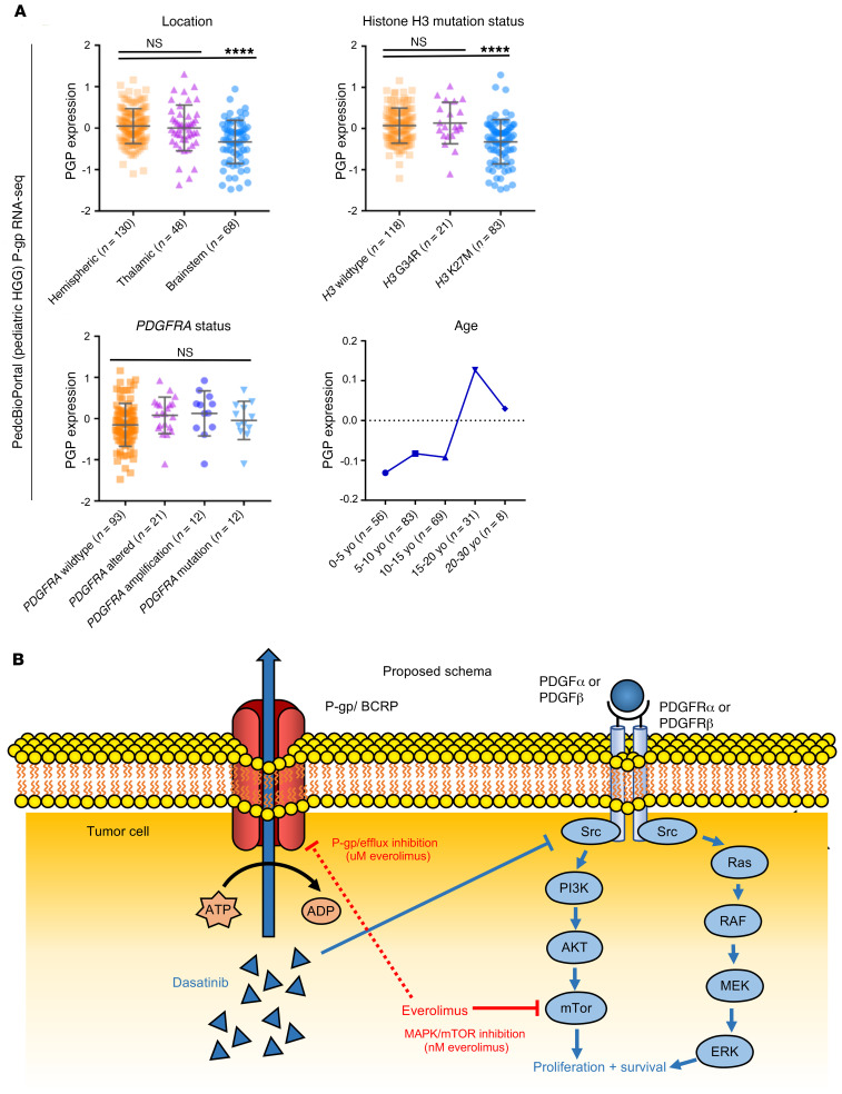
Pediatric and adult high-grade gliomas (HGGs) frequently harbor PDGFRA alterations. We hypothesized that cotreatment with everolimus may improve the efficacy of dasatinib in PDGFRα-driven glioma through combinatorial synergism and increased tumor accumulation of dasatinib. We performed dose-response, synergism, P-glycoprotein inhibition, and pharmacokinetic studies in in vitro and in vivo human and mouse models of HGG. Six patients with recurrent PDGFRα-driven glioma were treated with dasatinib and everolimus. We found that dasatinib effectively inhibited the proliferation of mouse and human primary HGG cells with a variety of PDGFRA alterations. Dasatinib exhibited synergy with everolimus in the treatment of HGG cells at low nanomolar concentrations of both agents, with a reduction in mTOR signaling that persisted after dasatinib treatment alone. Prolonged exposure to everolimus significantly improved the CNS retention of dasatinib and extended the survival of PPK tumor-bearing mice (mutant TP53, mutant PDGFRA, H3K27M). Six pediatric patients with glioma tolerated this combination without significant adverse events, and 4 patients with recurrent disease (n = 4) had a median overall survival of 8.5 months. Our results show that the efficacy of dasatinib treatment of PDGFRα-driven HGG was enhanced with everolimus and suggest a promising route for improving targeted therapy for this patient population.

Keywords: Brain cancer; Molecular biology; Oncology; Pharmacology.


Conflict of interest statement

Conflict of interest: AMC is on the Scientific Advisory Board of Tempus.

Figures

Figure 1
Figure 1. PDGFRα-driven glioma demonstrates sensitivity to dasatinib in vitro.
(A) HGG cells were generated from an IUE model with plasmids of TP53, PDGFRA, and H3K27M mutations. (B) Proliferation of IUE HGG cells in response to addition of PDGF α and β ligands in vitro (****P ≤ 0.0001, by Dunnett’s multiple comparisons test). n = 3 technical replicates. Avg., average. (C) Western blot analysis of p-PDGFRα and total PDGFRα in response to the addition of PDGF ligands. (D) Dose-response curve of PPK neurospheres, cultured cells from 6 human PDGFRα-driven glioma cell lines, and cells from 1 PDGFRA-WT glioma cell line following treatment with dasatinib. n = 3 technical replicates. (E) Plot of glioma cell culture sensitivity to dasatinib according to the GDSC database. Analysis of PDGFRA, PDGFA, PDGFRB, and PDGFB DNA alterations is referenced (#non-GDSC data generated from primary cell culture treatment for this study). Data represent the mean ± SEM for B and D.
Figure 2
Figure 2. Addition of everolimus provides synergism and strong P-gp blockade.
(A) Viability of IUE HGG neurospheres in response to various combinations of dasatinib and everolimus. n = 3 technical replicates. (B) Western blot analysis of p-PDGFRα/β (Tyr849/Tyr857), p-Src (Tyr416), and p-S6 (Ser235–236) expression in PDGFRA-amplified mouse HGG cells treated with dasatinib or everolimus monotherapy or with coadministration of dasatinib and everolimus. In the coadministration condition, the drugs were administered at equal doses (doses are indicated at the top of the figure). CTRL, control. (C) Quantification of Western blot analysis for the mouse HGG cell line (PPK) and a human PDGFRA-enhanced cell line (XIII-P), demonstrating a greater reduction in p-S6 expression with coadministration of dasatinib and everolimus than with either drug alone at 0.1 μM in the mouse cell line and at all administered doses in the human cell line. (D) A limiting dilution assay was performed on IUE PPK neurospheres treated with either 1 μM or 10 μM doses of dasatinib or everolimus, or dasatinib and everolimus together (5 μM). n = 3 technical replicates. (E) Everolimus and dasatinib synergism CI assessment. (F) Plot of P-gp inhibition using an in vitro assay with controls (higher numbers = greater P-gp inhibition) shows higher P-gp inhibition with the 1 μM everolimus dose condition than with the negative control or no treatment conditions ( **P = 0.0045 and ****P ≤ 0.0001, by Tukey’s multiple comparisons test, respectively). Neg., negative; Pos., positive. Data represent the mean ± SEM (A, D, and F).
Figure 3
Figure 3. Sustained exposure of everolimus improves dasatinib tumor concentration and efficacy in a mouse model.
(A) PK data documenting an increase in dasatinib levels detected in tumors with an additional 24-hour everolimus pre-dasatinib dose of either 5 mg/kg or 10 mg/kg (*P ≤ 0.05, by Dunnett’s multiple comparisons test). n = 5 replicates. (B) Survival curve data for mice with PDGFRA-amplified HGG via IUE shows that the median survival for the control condition was 52 days after IUE injection (n = 10), 55 days for the everolimus condition (5 mg/kg, n = 9), 61 days for the dasatinib condition (10 mg/kg, n = 9), and 86 days for the dasatinib plus everolimus condition (10 mg/kg and 5 mg/kg, respectively, n = 9) (NS P = 0.3378, ***P ≤ 0.0005, and ****P ≤ 0.0001, by log-rank test). (C) Representative bioluminescence images of PPK mouse tumors (vehicle, n = 10; dasatinib, n = 9; everolimus, n = 9; dasatinib + everolimus, n = 10) 47 days after IUE injection, displaying lower average luminescence in the cotreatment group than in the 2 groups receiving monotherapy of dasatinib or everolimus. (D) IUE PPK bioluminescence tumor monitor growth data with statistical significance between dasatinib plus everolimus and DMSO treatment groups 47 days after IUE injection (****P ≤ 0.0001, by Tukey’s multiple comparisons test.). (E) In vivo IHC images of p-Src and p-S6 expression levels, documenting a reduction with everolimus and dasatinib treatment in comparison with nontreated tumors. Images are representative of 3 biological replicates. Original magnification, ×20. (F) IHC quantification data for treated PPK mice show statistical significance of p-Src and p-S6 expression levels between tumors treated with dasatinib and everolimus and nontreated tumors (**P = 0.0065 and ****P ≤ 0.0001, by Dunnett’s multiple comparisons test). n = 3 animals per treatment group and 4 images per animal. Data represent the mean ± SEM (A, D, and F).
Figure 4
Figure 4. Patient survival curves and treatment of UMPED44 with relapsed PDGFRA-amplified HGG.
(A) PFS curve for patients with recurrent PDGFRα-driven HGG treated with dasatinib and everolimus (n = 4). Dashed lines represent a 95% confidence interval. (B) OS curve for patients with recurrent PDGFRα-driven HGG treated with dasatinib and everolimus (n = 4). Dashed lines represent a 95% confidence interval. (C) UMPED44 axial FLAIR MRI at baseline, before cycle 3, and before cycle 4 (MR spectroscopy in cycle 4 confirmed tumor progression).
Figure 5
Figure 5. P-gp expression in human pediatric HGG and proposed schema.
(A) PedcBioPortal (pediatric HGG) P-gp RNA-Seq data by location (upper left), histone H3 mutation status (upper right), PDGFRA status (lower left), and age (lower right) (****P ≤ 0.0001, by Tukey’s multiple comparisons test). Data presented in the upper 2 graphs and lower left graph reflect the mean ± SEM. (B) Proposed schema of dasatinib and everolimus targeting of PDGF-driven HGG. Red line depicting inhibition of P-gp/BCRP by everolimus is dashed to reflect the higher concentration (μM) required to achieve P-gp inhibition than to achieve MAPK/mTOR inhibition (nM). yo, years old.

References

    1. Liu J, et al. An integrated TCGA Pan-cancer clinical data resource to drive high-quality survival outcome analytics. Cell. 2018;173(2):400–416.e11. doi: 10.1016/j.cell.2018.02.052. - DOI - PMC - PubMed
    1. Mackay A, et al. Integrated molecular meta-analysis of 1,000 pediatric high-grade and diffuse intrinsic pontine glioma. Cancer Cell. 2017;32(4):520–537.e5. doi: 10.1016/j.ccell.2017.08.017. - DOI - PMC - PubMed
    1. Farahani RM, Xaymardan M. Platelet-derived growth factor receptor alpha as a marker of mesenchymal stem cells in development and stem cell biology. Stem Cells Int. 2015;2015:362753. - PMC - PubMed
    1. Mackay A, et al. Molecular, pathological, radiological, and immune profiling of non-brainstem pediatric high-grade glioma from the HERBY phase II randomized trial. Cancer Cell. 2018;33(5):829–842.e5. doi: 10.1016/j.ccell.2018.04.004. - DOI - PMC - PubMed
    1. Koschmann C, et al. Characterizing and targeting PDGFRA alterations in pediatric high-grade glioma. Oncotarget. 2016;7(40):65696–65706. doi: 10.18632/oncotarget.11602. - DOI - PMC - PubMed

Publication types

MeSH terms

Substances