

Massive Release of CD9+ Microvesicles in Human Immunodeficiency Virus Infection, Regardless of Virologic Control
- PMID: 32603406
- PMCID: PMC8922002
- DOI: 10.1093/infdis/jiaa375
Massive Release of CD9+ Microvesicles in Human Immunodeficiency Virus Infection, Regardless of Virologic Control
Abstract
Background: The role of extracellular vesicles (EVs) in human immunodeficiency virus (HIV) pathogenesis is unknown. We examine the cellular origin of plasma microvesicles (MVs), a type of ectocytosis-derived EV, the presence of mitochondria in MVs, and their relationship to circulating cell-free mitochondrial deoxyribonucleic acid (ccf-mtDNA) in HIV-infected patients and controls.
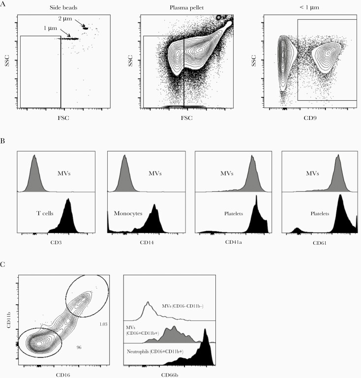
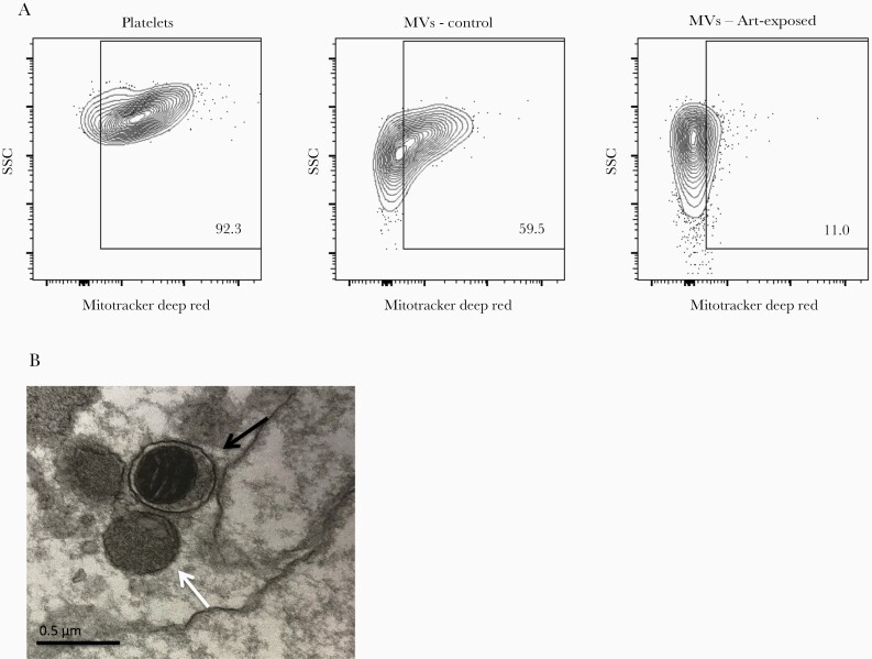
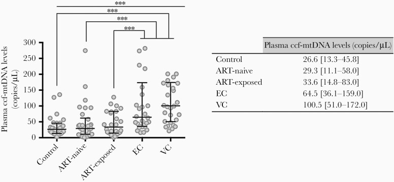
Methods: Five participant groups were defined: 30 antiretroviral therapy (ART)-naive; 30 ART-treated with nondetectable viremia; 30 elite controllers; 30 viremic controllers; and 30 HIV-uninfected controls. Microvesicles were quantified and characterized from plasma samples by flow cytometry. MitoTrackerDeepRed identified MVs containing mitochondria and ccf-mtDNA was quantified by real-time polymerase chain reaction.
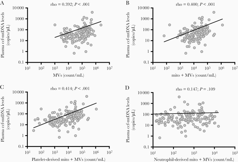
Results: Microvesicle numbers were expanded at least 10-fold in all HIV-infected groups compared with controls. More than 79% were platelet-derived MVs. Proportions of MVs containing mitochondria (22.3% vs 41.6%) and MV mitochondrial density (706 vs 1346) were significantly lower among HIV-infected subjects than controls, lowest levels for those on ART. Microvesicle numbers correlated with ccf-mtDNA levels that were higher among HIV-infected patients.
Conclusions: A massive release of platelet-derived MVs occurs during HIV infection. Some MVs contain mitochondria, but their proportion and mitochondrial densities were lower in HIV infection than in controls. Platelet-derived MVs may be biomarkers of platelet activation, possibly reflecting pathogenesis even in absence of HIV replication.
Keywords: HIV; elite controllers; extracellular vesicles; microvesicles; mitochondria.
© The Author(s) 2020. Published by Oxford University Press for the Infectious Diseases Society of America.
Figures
References
-
- Palella FJ Jr, Delaney KM, Moorman AC, et al. . Declining morbidity and mortality among patients with advanced human immunodeficiency virus infection. HIV Outpatient Study Investigators. N Engl J Med 1998; 338:853–60. - PubMed
-
- French MA, King MS, Tschampa JM, da Silva BA, Landay AL. Serum immune activation markers are persistently increased in patients with HIV infection after 6 years of antiretroviral therapy despite suppression of viral replication and reconstitution of CD4+ T cells. J Infect Dis 2009; 200:1212–5. - PubMed
