. 2020 Aug 14;6(8):2086-2098.
doi: 10.1021/acsinfecdis.0c00125. Epub 2020 Jul 14.

Evaluation of Peptide-Based Probes toward In Vivo Diagnostic Imaging of Bacterial Biofilm-Associated Infections

Affiliations

Evaluation of Peptide-Based Probes toward In Vivo Diagnostic Imaging of Bacterial Biofilm-Associated Infections

Landon W Locke et al. ACS Infect Dis. .

Abstract

The clinical management of bacterial biofilm infections represents an enormous challenge in today's healthcare setting. The NIH estimates that 65% of bacterial infections are biofilm-related, and therapeutic outcomes are positively correlated with early intervention. Currently, there is no reliable imaging technique to detect biofilm infections in vivo, and current clinical protocols for accurate and direct biofilm identification are nonexistent. In orthopedic implant-associated biofilm infections, for example, current detection methods are based on nonspecific X-ray or radiolabeled white blood cell imaging, coupled with peri-prosthetic tissue or fluid samples taken invasively, and must be cultured. This approach is time-consuming and often fails to detect biofilm bacteria due to sampling errors and a lack of sensitivity. The ability to quantify bacterial biofilms by real-time noninvasive imaging is an urgent unmet clinical need that would revolutionize the management and treatment of these devastating types of infections. In the present study, we assembled a collection of fluorescently labeled peptide candidates to specifically explore their biofilm targeting properties. We evaluated these fluorescently labeled peptides using various in vitro assays for their ability to specifically and nondestructively target biofilms produced by model bacterial pathogen Pseudomonas aeruginosa. The lead candidate that emerged, 4Iphf-HN17, demonstrated rapid biofilm labeling kinetics, a lack of bactericidal activity, and biofilm targeting specificity in human cell infection models. In vivo fluorescently labeled 4Iphf-HN17 showed enhanced accumulation in biofilm-infected wounds, thus warranting further study.

Keywords: Pseudomonas aeruginosa; biofilms; diagnostics; imaging; optical; peptides.


Figures

Figure 1.
Figure 1.
Specific detection of bacterial biofilms by targeted probes that can be localized in the body by imaging. Following probe administration, non-invasive in vivo imaging allows the infection site to be identified and localized. Cartoon on the right shows the labeled probe recognizing and accumulating in biofilms surface-attached to eukaryotic cells.
Figure 2.
Figure 2.
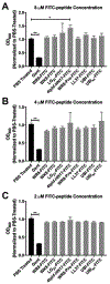
Normalized OD600 measurements of P. aeruginosa cultures following 16 h exposure to FITC-labeled peptides applied at 8 μM (A), 4 μM (B), and 2 μM (C). Data are presented as the fold change in OD600 compared to PBS-treated P. aeruginosa cultures. The FITC-labeled peptide candidates do not affect growth of P. aeruginosa, with the exception of 4Iphf-HN17-FITC applied at applied at the highest concentration (8 μM). The positive control gentamicin significantly inhibited growth as expected. Statistical significance was determined by one-way ANOVA followed by Dunnett’s multiple-comparison test. Data derive from two independent experiments and are presented as mean ± SEM. **p < 0.0001 and *p < 0.01.
Figure 3.
Figure 3.
Representative CLSM images showing FITC-peptide labeling of flow cell-generated P. aeruginosa biofilms. The biofilms were grown for 24 h and treated with FITC-peptides at 2 μM. The green and red channels show FITC-peptide and FM4-64 staining, respectively. The images show a single slice through the biofilms at mid-height of the structures. Scale bar = 20 μm. FITC-peptides that localized to the periphery of biofilms are indicated by arrow heads while FITC-peptides that penetrated biofilms are indicated by arrows.
Figure 4.
Figure 4.
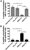
Representative CLSM images used for evaluating FITC-peptide distribution in a co-culture model of epithelial cells with mCherry-expressing P. aeruginosa. 4Iphf-HN17-FITC and LG21-FITC demonstrated selectivity for bacteria while other probes showed minimal bacterial targeting or showed targeting to both bacteria and epithelial cells. Each panel shows a representative image of at least two independent experiments for each peptide, with >8 images acquired per experiment. Epithelial A549 cells were prelabeled with cell tracker blue (CTB) for visualization. Scale bar = 20 μm.
Figure 5.
Figure 5.
CLSM image colocalization analysis of select FITC-labeled peptides. Probe colocalization is reported as the % colocalization with epithelial cells (A) or bacteria (B) normalized by total detected pixel area of the probe. Data shown are combined from two separate experiments and error bars represent SEM. FITC-probes that showed no detectable signal above a background threshold (LL37-FITC, WR8-pro-FITC, UBI29-41-FITC, and UBIsc-FITC) were excluded from analysis. *p < 0.0003 and **p = 0.001.
Figure 6.
Figure 6.
4Iphf-HN17-Cy5 preferentially labels P. aeruginosa-infected human neutrophils in vitro. Panels show representative images of (A) neutrophils infected with PAO1-GFP+ in the absence of probe, (B) neutrophils incubated with probe in the absence of infection, and (C) infected neutrophils incubated with 4Iphf-HN17-Cy5. Images are from one representative donor. Scale bar = 7 μm. (D) Normalized Cy5 mean fluorescence intensity (MFI) on four different neutrophil populations based on infection status and neutrophil morphology. Data shown are from two different donors and error bars represent the standard deviation of the means. *p = 0.008.
Figure 7.
Figure 7.
In vivo optical imaging evaluation of 4Iphf-HN17-Cy5 in a mouse model of wound infection. Panels show bioluminescence and epi fluorescence images of representative mice from each group detecting lux-tagged P. aeruginosa and Cy5 distribution, respectively. (A) Mouse with P. aeruginosa-infected wounds injected with 4Iphf-HN17-Cy5, (B) mouse with PBS-treated wounds injected with 4Iphf-HN17-Cy5, and (C) mouse with lux-tagged P. aeruginosa-infected wounds injected with PBS. All images were acquired 18 h after probe injection. The min/max scale was set equally for all images. (D) ROI-derived Cy5 wound fluorescence normalized to surrounding non-wound tissue 18 h after injection for the three study groups. *p < 0.001.
Figure 8.
Figure 8.
In situ CLSM images of 4Iphf-HN17-Cy5 distribution in wound tissue. Representative confocal micrographs of tissue sections of 4Iphf-HN17-Cy5 injected mice with (A) PBS-treated wounds and (B) P. aeruginosa-infected wounds. The images revealed a punctate localization pattern of 4Iphf-HN17-Cy5 in areas immediately adjacent to TdTomato-positive P. aeruginosa cells and multi-cellular aggregates. Arrowheads indicate cell-shaped localization patterns of Cy5 fluorescence near pockets of P. aeruginosa aggregates. Scale bar = 10 μm.

References

    1. Davies D, Understanding biofilm resistance to antibacterial agents. Nat Rev Drug Discov 2003, 2 (2), 114–22. - PubMed
    1. Bjarnsholt T; Alhede M; Alhede M; Eickhardt-Sorensen SR; Moser C; Kuhl M; Jensen PO; Hoiby N, The in vivo biofilm. Trends Microbiol 2013, 21 (9), 466–74. - PubMed
    1. Flemming HC; Wingender J, The biofilm matrix. Nat Rev Microbiol 2010, 8 (9), 623–33. - PubMed
    1. Joo HS; Otto M, Molecular basis of in vivo biofilm formation by bacterial pathogens. Chem Biol 2012, 19 (12), 1503–13. - PMC - PubMed
    1. Bjarnsholt T, The role of bacterial biofilms in chronic infections. APMIS Suppl 2013, (136), 1–51. - PubMed

Publication types