

The Effect of Tube Location on Corneal Endothelial Cells in Patients with Ahmed Glaucoma Valve
- PMID: 32603727
- PMCID: PMC7765742
- DOI: 10.1016/j.ophtha.2020.06.050
The Effect of Tube Location on Corneal Endothelial Cells in Patients with Ahmed Glaucoma Valve
Abstract
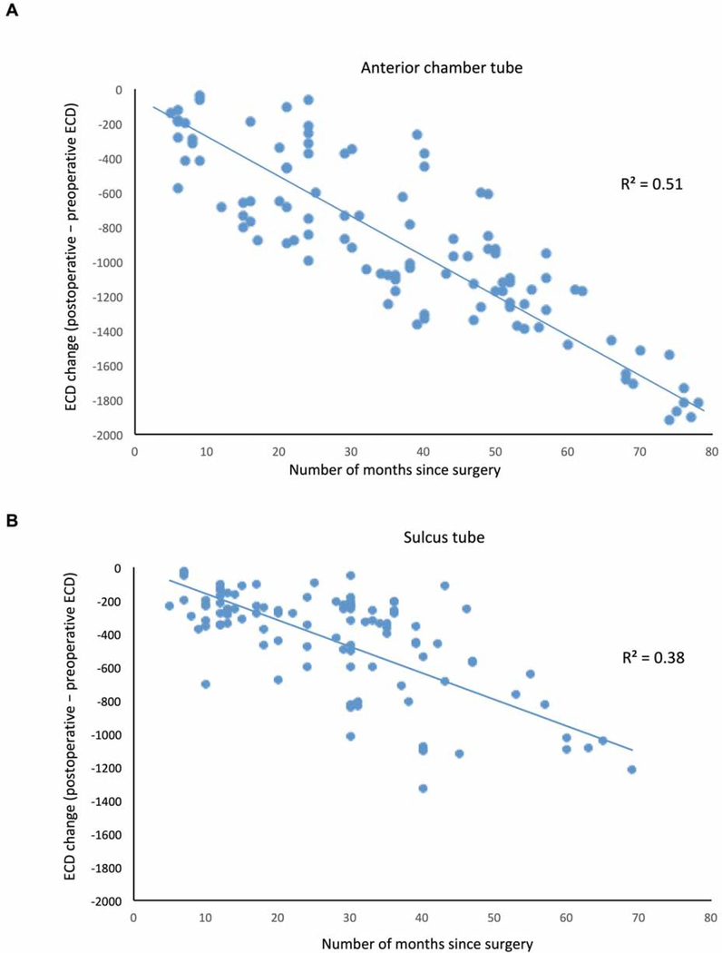
Purpose: To compare the effects of the Ahmed glaucoma valve (AGV; New World Medical, Rancho Cucamonga, CA) with sulcus versus anterior chamber (AC) tube placement on the corneal endothelial density and morphology over time.
Design: Nonrandomized, interventional study.
Participants: This study included 106 eyes from 101 pseudophakic patients who had the AGV tube placed in the AC (acAGV) and 105 eyes from 94 pseudophakic patients who had the AGV tube placed in the ciliary sulcus (sAGV).
Methods: All patients underwent preoperative specular microscopy, which was repeated postoperatively in 2019. The patients' demographic information, glaucoma diagnoses, and basic ocular information were obtained on chart review. Anterior segment OCT was conducted for patients who underwent sAGV to evaluate the sulcus tube position. Gonioscopy was performed to document peripheral anterior synechiae (PAS). Linear mixed-effects models were used to compare the different ocular and endothelial measurements between the 2 groups and to identify risk factors for endothelial cell density (ECD) loss over time.
Main outcome measures: Monthly change in corneal endothelial measurements, including ECD and coefficient of variation (CV), calculated as the difference between preoperative and postoperative measurements divided by the number of months from the time of surgery to postoperative specular microscopy.
Results: The acAGV and sAGV groups were comparable in all baseline characteristics except that the acAGV group had longer follow-up (37.6 vs. 20.1 months, respectively, P < 0.001). Mean monthly loss in central ECD was significantly more in the acAGV group (mean ± standard deviation: 29.3±29.7 cells/mm2) than in the sAGV group (15.3±20.7 cells/mm2, P < 0.0001). Mean monthly change in CV was similar between the 2 groups (P = 0.28). Multivariate analyses revealed that younger age and tube location in the AC were associated with faster central ECD loss (P = 0.02, P < 0.0001, respectively). For patients with sAGV, while PAS was associated with faster central ECD loss (P = 0.002), a more forward tube position tenting the iris was not (P > 0.05).
Conclusions: Compared with anterior segment placement, ciliary sulcus tube implantation may be a preferred surgery approach to reduce endothelial cell loss in pseudophakic patients.
Published by Elsevier Inc.
Conflict of interest statement
Conflict of interest: No conflicting relationship exists for any author.
Figures

References
-
- Arora KS, Robin AL, Corcoran KJ, Corcoran SL, Ramulu PY. Use of Various Glaucoma Surgeries and Procedures in Medicare Beneficiaries from 1994 to 2012. Ophthalmology. 2015;122(8):1615–1624, - PubMed
-
- Minckler DS, Francis BA, Hodapp EA, et al. Aqueous shunts in glaucoma: a report by the American Academy of Ophthalmology. Ophthalmology. 2008;115(6):1089–1098. - PubMed
-
- Hau S, Scott A, Bunce C, Barton K. Corneal endothelial morphology in eyes implanted with anterior chamber aqueous shunts. Cornea. 2011;30(1):50–55. - PubMed
Publication types
MeSH terms
Grants and funding
LinkOut - more resources
Full Text Sources
Other Literature Sources
