

SARS-CoV-2 pathophysiology and assessment of coronaviruses in CNS diseases with a focus on therapeutic targets
- PMID: 32603829
- PMCID: PMC7320676
- DOI: 10.1016/j.bbadis.2020.165889
SARS-CoV-2 pathophysiology and assessment of coronaviruses in CNS diseases with a focus on therapeutic targets
Abstract
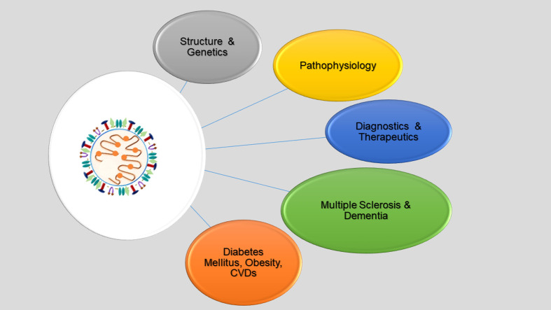
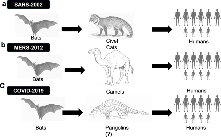
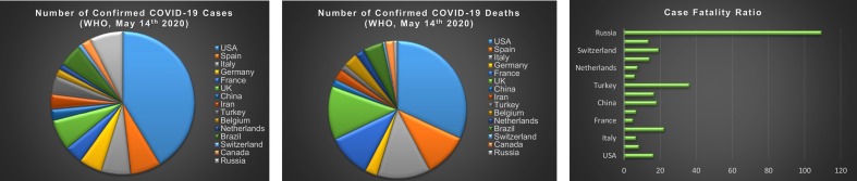
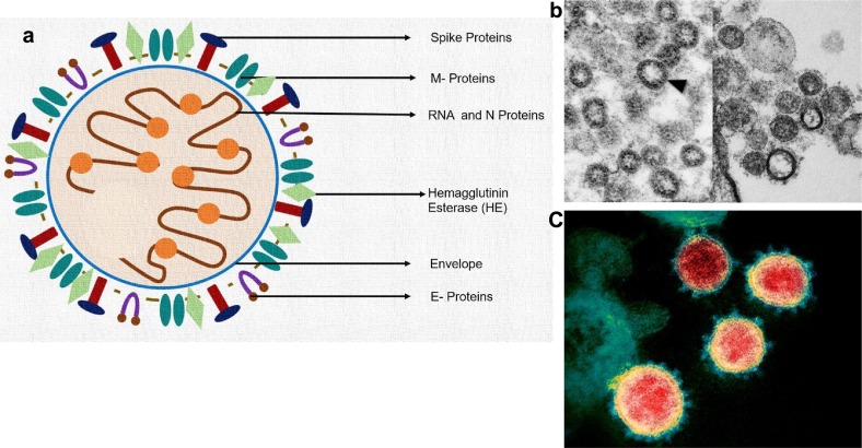
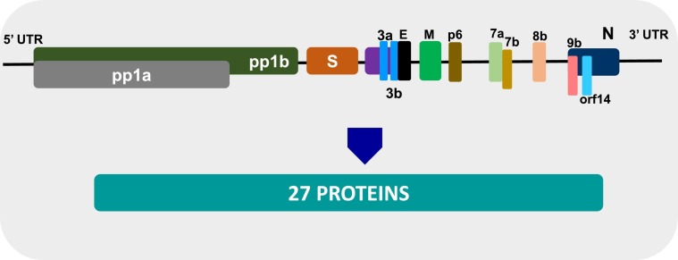
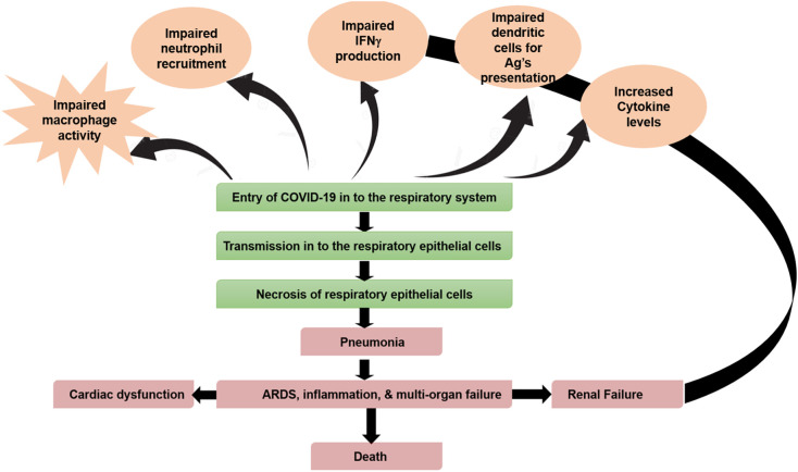
The novel Coronavirus disease of 2019 (nCOV-19) is a viral outbreak noted first in Wuhan, China. This disease is caused by Severe Acute Respiratory Syndrome (SARS) Coronavirus (CoV)-2. In the past, other members of the coronavirus family, such as SARS and Middle East Respiratory Syndrome (MERS), have made an impact in China and the Arabian peninsula respectively. Both SARS and COVID-19 share similar symptoms such as fever, cough, and difficulty in breathing that can become fatal in later stages. However, SARS and MERS infections were epidemic diseases constrained to limited regions. By March 2020 the SARS-CoV-2 had spread across the globe and on March 11th, 2020 the World Health Organization (WHO) declared COVID-19 as pandemic disease. In severe SARS-CoV-2 infection, many patients succumbed to pneumonia. Higher rates of deaths were seen in older patients who had co-morbidities such as diabetes mellitus, hypertension, cardiovascular disease (CVD), and dementia. In this review paper, we discuss the effect of SARS-CoV-2 on CNS diseases, such as Alzheimer's-like dementia, and diabetes mellitus. We also focus on the virus genome, pathophysiology, theranostics, and autophagy mechanisms. We will assess the multiorgan failure reported in advanced stages of SARS-CoV-2 infection. Our paper will provide mechanistic clues and therapeutic targets for physicians and investigators to combat COVID-19.
Keywords: Brain; COVID-19; Diabetes mellitus; Multiple sclerosis; Neutralizing antibodies; SARS-CoV-2; Therapeutics.
Copyright © 2020 Elsevier B.V. All rights reserved.
Conflict of interest statement
Declaration of competing interest The authors declare that they have no known competing financial interests or personal relationships that could have appeared to influence the work reported in this paper.
Figures
Similar articles
-
3C-like protease inhibitors block coronavirus replication in vitro and improve survival in MERS-CoV-infected mice.Sci Transl Med. 2020 Aug 19;12(557):eabc5332. doi: 10.1126/scitranslmed.abc5332. Epub 2020 Aug 3. Sci Transl Med. 2020. PMID: 32747425 Free PMC article.
-
COVID-19: a conundrum to decipher.Eur Rev Med Pharmacol Sci. 2020 May;24(10):5830-5841. doi: 10.26355/eurrev_202005_21378. Eur Rev Med Pharmacol Sci. 2020. PMID: 32495923
-
The origin, transmission and clinical therapies on coronavirus disease 2019 (COVID-19) outbreak - an update on the status.Mil Med Res. 2020 Mar 13;7(1):11. doi: 10.1186/s40779-020-00240-0. Mil Med Res. 2020. PMID: 32169119 Free PMC article. Review.
-
Remdesivir against COVID-19 and Other Viral Diseases.Clin Microbiol Rev. 2020 Oct 14;34(1):e00162-20. doi: 10.1128/CMR.00162-20. Print 2020 Dec 16. Clin Microbiol Rev. 2020. PMID: 33055231 Free PMC article. Review.
-
Severe acute respiratory syndrome coronavirus-2 (SARS-CoV-2), a newly emerged pathogen: an overview.Pathog Dis. 2020 Aug 1;78(6):ftaa042. doi: 10.1093/femspd/ftaa042. Pathog Dis. 2020. PMID: 32840560 Free PMC article. Review.
Cited by
-
A Comparative NLP-Based Study on the Current Trends and Future Directions in COVID-19 Research.IEEE Access. 2021 May 20;9:78341-78355. doi: 10.1109/ACCESS.2021.3082108. eCollection 2021. IEEE Access. 2021. PMID: 34786315 Free PMC article.
-
Orthotopic Liver Transplantation of a SARS-CoV-2 Negative Recipient from a Positive Donor: The Border between Uncertainty and Necessity in a Pandemic Era- Case Report and Overview of the Literature.Medicina (Kaunas). 2023 Apr 26;59(5):836. doi: 10.3390/medicina59050836. Medicina (Kaunas). 2023. PMID: 37241068 Free PMC article. Review.
-
Computational repurposing of tamibarotene against triple mutant variant of SARS-CoV-2.Comput Biol Med. 2021 Sep;136:104748. doi: 10.1016/j.compbiomed.2021.104748. Epub 2021 Aug 8. Comput Biol Med. 2021. PMID: 34388463 Free PMC article.
-
Possible therapeutic targets and promising drugs based on unsymmetrical hetaryl-substituted porphyrins to combat SARS-CoV-2.J Pharm Anal. 2021 Dec;11(6):691-698. doi: 10.1016/j.jpha.2021.08.003. Epub 2021 Aug 5. J Pharm Anal. 2021. PMID: 34377564 Free PMC article.
-
Long-Term Respiratory and Neurological Sequelae of COVID-19.Med Sci Monit. 2020 Nov 1;26:e928996. doi: 10.12659/MSM.928996. Med Sci Monit. 2020. PMID: 33177481 Free PMC article. Review.
References
-
- Enquist L.W., Dermody T.S., DiMaio D. Vol. 3. 2016. Introduction, Annual Review of Virology. v. - PubMed
-
- Beachy R.N., Zaitlin M. Replication of tobacco mosiac virus, VI Replicative intermediate and TMV-RNA-related RNAs associated with polyribosomes. Virology. 1975;63:84–97. - PubMed
-
- Reddi K.K. Degradation of tobacco mosiac virus nucleic acid with micrococcal phosphodiesterase. Biochim. Biophys. Acta. 1959;36:132–142. - PubMed
-
- Hart R.G. Infectivity measurements of partially degraded tobacco mosiac virus. Virology. 1955;1:402–407. - PubMed
Publication types
MeSH terms
Substances
Grants and funding
LinkOut - more resources
Full Text Sources
Miscellaneous
