Observational Study
. 2021 Feb 1;41(2):381-386.
doi: 10.1097/IAE.0000000000002875.

FINGERPRINT SIGN OF THE HENLE FIBER LAYER

Affiliations
Observational Study

FINGERPRINT SIGN OF THE HENLE FIBER LAYER

Shane M Griffin et al. Retina. .

Abstract

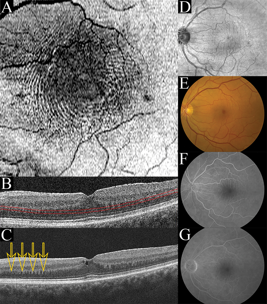
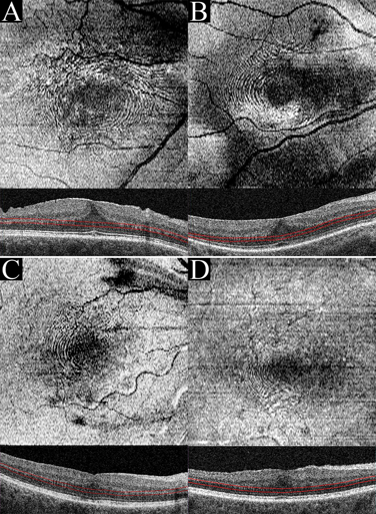
Purpose: To describe the appearance of concentric, fingerprint-like waves within the Henle fiber layer (HFL) using en face optical coherence tomography in patients with tractional pathologies of the retina.

Methods: Retrospective analysis of six eyes of six patients imaged by optical coherence tomography with volumetric slabs positioned at the level of the HFL.

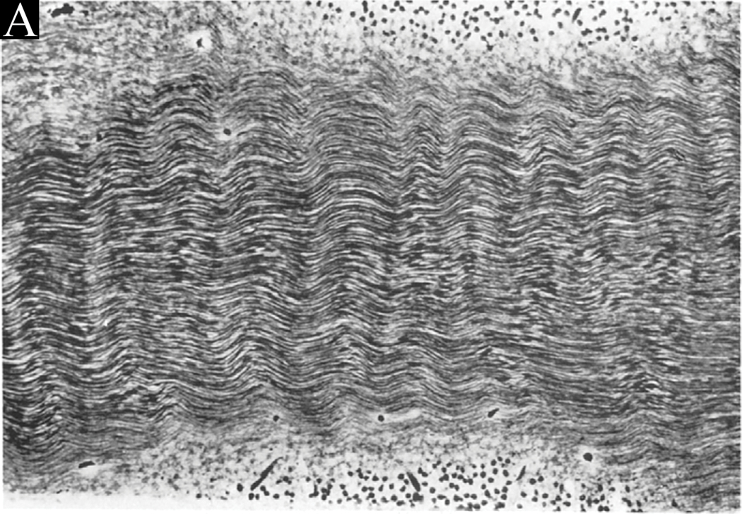
Results: Optical coherence tomography data from six patients with tractional vitreoretinal pathology were reviewed. Concentric, fingerprint-like microwaves were visualized through en face optical coherence tomography in all six study eyes at the level of the HFL. This finding resembled the finding of HFL waves previously noted histopathologically from force exerted on this layer.

Conclusion: In retinal pathologies in which specific physical forces act on the retina, volumetric optical coherence tomography may permit visualization of en face concentric, fingerprint-like hyperreflective rings within the HFL. This "fingerprint sign" may represent a biomechanical consequence of traction on the retina and allow clinical decision making based on improved recognition of the existence of such traction.


Figures

Figure 1:
Figure 1:
(Case 1): En face concentric waves (A) are seen when a contoured slab is positioned at the level of the HFL (B). Averaged B-scan through the fovea (C) reveals a dentate appearance of the synaptic OPL/HFL junction (arrows) and a hyper-reflective ERM is seen. Infrared imaging (D), Fundus photography (E), early fluorescein angiography (F), and late fluorescein angiography (G) do not show the concentric wave pattern.
Figure 2:
Figure 2:
When RPE-contoured slabs of thickness between 31–47 microns were positioned at the level of the HFL, the en face “fingerprint sign” was visualized. Case 2(A), Case 3(B), Case 4(C), and Case 5(D) all presented with ERM.
Figure 3:
Figure 3:
Wolter’s histological photographs demonstrating physical waves within the HFL. “Fig. 4. (Wolter) A part of the temporal aspect of Henle’s fiber layer exhibiting regular ‘microwaves’ with ridges and valleys in a vertical direction. All fibers are well preserved. Flat frozen section, Hortega stain, photomicrograph × 300”

References

    1. Curcio CA, Allen KA. Topography of ganglion cells in human retina. J Comp Neurol. 1990;300(1):5–25. doi:10.1002/cne.903000103 - DOI - PubMed
    1. Govetto A, Hubschman J-P, Sarraf D, et al. The role of Müller cells in tractional macular disorders: an optical coherence tomography study and physical model of mechanical force transmission. Br J Ophthalmol. 2019:bjophthalmol-2019–314245. doi:10.1136/bjophthalmol-2019-314245 - DOI - PubMed
    1. Pang CE, Spaide RF, Freund KB. Epiretinal proliferation seen in association with lamellar macular holes: A distinct clinical entity. Retina. 2014;34(8):1513–1523. doi:10.1097/IAE.0000000000000163 - DOI - PubMed
    1. Lujan BJ, Roorda A, Knighton RW, Carroll J. Revealing Henle’s fiber layer using spectral domain optical coherence tomography. Investig Ophthalmol Vis Sci. 2011;52(3):1486–1492. doi:10.1167/iovs.10-5946 - DOI - PMC - PubMed
    1. Ouyang Y, Walsh AC, Keane PA, et al. Different phenotypes of the appearance of the outer plexiform layer on optical coherence tomography. Graefe’s Arch Clin Exp Ophthalmol. 2013;251(10):2311–2317. doi:10.1007/s00417-013-2308-5 - DOI - PubMed

Publication types